Coloured SEM of anastomosis of femoral artery
Numéro d’image : 11840144
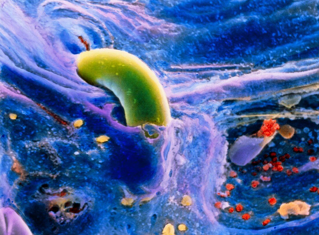
| Arterial anastomosis. Coloured scanning electron micrograph of the lumen of the femoral artery showing its internal wall,following and end to end anastomosis. This is a surgical procedure in which the artery is cut and part of it is removed; the two healthy ends of the artery are then sutured (green) together. It is carried out to remove regions of an artery which have become blocked due to thrombi (clots) or atheroma (fat) plaques. Some red blood cells are seen at bottom right and platelets (yellow) are at bottom. Magnification: x3100 at 6x7cm size | |
| Licence : | Droits gérés |
| Crédit: | Science Photo Library / PROFESSORS P.M. MOTTA, G. MACCHIARELLI, A. CAGGIATI & F.M. MAGLIOCCA |
| Taille de l’image : | 4919 px × 3626 px |
| Model Release : | Non requis |
| Property Release : | Non requis |
| Restrictions : | - |
Prix pour cette image À partir de 45 €
Produit vendu
(Calendrier, Carte postale, Carte de vœux, Impression sur textile, Packaging etc)
À partir de 45 €
Usage commercial
(Affichage, Annonce presse, Annonce TV, Carte, Digital - hors rés. sociaux, Digital - rés. sociaux etc)
À partir de 45 €
Éditorial
(Digital, Journal, Livre, Livre pratique, Magazine, Télévision etc)
À partir de 60 €
Usage non-commercial
(Digital - hors rés. sociaux, Digital - rés. sociaux etc)
À partir de 120 €