Light micrograph of cardiac muscle post infarction
Numéro d’image : 11839762
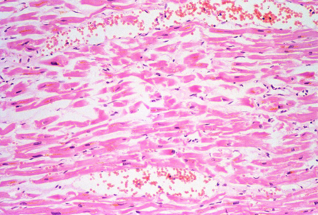
| Light micrograph of human cardiac muscle following infarction. In longitudinal section,the muscle fibres (lilac) run horizontally. Abnormal features are loss of organisation in the fibres,reduced numbers of nuclei (purple) and increased space (white) between the fibres,representing interstitial oedema. The small round cells stained red at top and bottom are neutrophils infiltrating the oedematous interstitium. Together with macrophages,neutrophils represent an early inflammatory response; they serve to remove the necrotic tissue prior to scar formation. Magnification x50 at 35mm size | |
| Licence : | Droits gérés |
| Crédit: | Science Photo Library / Michler, Astrid & Hans-Frieder |
| Taille de l’image : | 5230 px × 3543 px |
| Model Release : | Non requis |
| Property Release : | Non requis |
| Restrictions : | - |
Prix pour cette image À partir de 45 €
Produit vendu
(Calendrier, Carte postale, Carte de vœux, Impression sur textile, Packaging etc)
À partir de 45 €
Usage commercial
(Affichage, Annonce presse, Annonce TV, Carte, Digital - hors rés. sociaux, Digital - rés. sociaux etc)
À partir de 45 €
Éditorial
(Digital, Journal, Livre, Livre pratique, Magazine, Télévision etc)
À partir de 60 €
Usage non-commercial
(Digital - hors rés. sociaux, Digital - rés. sociaux etc)
À partir de 120 €