Collapsed lung,CT scan
Numéro d’image : 11839611
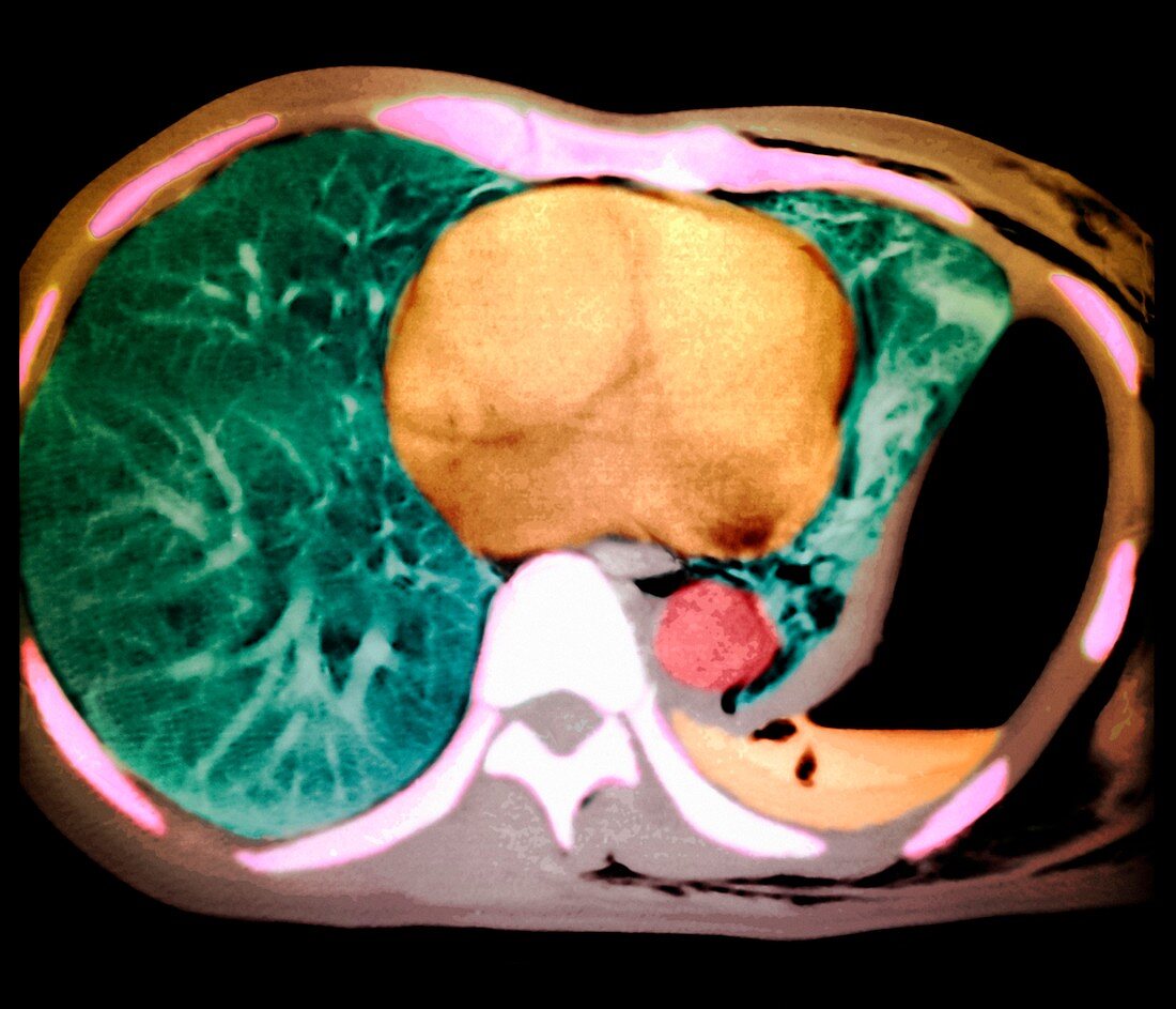
| Collapsed lung. Coloured computed tomography (CT) scan through a patient's chest. The front of the body is at top. A pneumothorax,a large pocket of air (black,centre right to lower right),has expanded the pleural cavity and compressed the patient's left lung (green). The right lung (left) is not compressed,because the pleural cavity has its normal shape of a thin space between the lungs and the chest wall. The heart (orange,centre) is between the lungs. Fluid (orange,lower right) has gathered in the pneumothorax,a complication named a hydropneumothorax. A splashing sound is heard if the patient is shaken. Surgical insertion of a chest tube is needed to drain the air and fluid | |
| Licence : | Droits gérés |
| Crédit: | Science Photo Library / Zephyr |
| Taille de l’image : | 4134 px × 3543 px |
| Model Release : | Non requis |
| Property Release : | Non requis |
| Restrictions : | - |
Prix pour cette image À partir de 45 €
Produit vendu
(Calendrier, Carte postale, Carte de vœux, Impression sur textile, Packaging etc)
À partir de 45 €
Usage commercial
(Affichage, Annonce presse, Annonce TV, Carte, Digital - hors rés. sociaux, Digital - rés. sociaux etc)
À partir de 45 €
Éditorial
(Digital, Journal, Livre, Livre pratique, Magazine, Télévision etc)
À partir de 60 €
Usage non-commercial
(Digital - hors rés. sociaux, Digital - rés. sociaux etc)
À partir de 120 €
Mots clés
- axial,
- buste,
- cage thoracique,
- catégorie,
- coloré,
- colorié,
- colorisé,
- coupe,
- désordre,
- diagnostic,
- diagnostique,
- divisé,
- effondré,
- état,
- fluide,
- maladie,
- médecine,
- médical,
- médicale,
- partie,
- patient,
- patients,
- pneumothorax,
- poche d'air,
- poumon,
- pulmonaire,
- respiration,
- respiratoire,
- section,
- soins de santé,
- thoracique,
- thorax,
- tomodensitométrie,
- tomographie assistée par ordinateur,
- trouble