X-ray image showing gallbladder with gall
Numéro d’image : 11839319
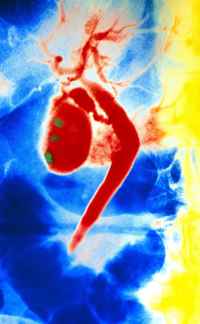
| Coloured X-ray showing gallstones within the gall bladder (red,pear-shaped). Gallstones,hard masses formed by bile pigments,cholesterol and calcium salts,are seen as green areas in the gall bladder. The technique used to obtain this image is known as endoscopic cholangiography. The contrast medium is injected into the common bile duct (bent tube at centre) with an endoscope which is passed via the mouth,stomach and duodenum. Gallstones that do not cause symptoms may be safely left untreated. If the symptoms are severe a cholecystectomy,the surgical removal of the gallbladder,may be carried out | |
| Licence : | Droits gérés |
| Crédit: | Science Photo Library / DEPT. OF CLINICAL RADIOLOGY, SALISBURY DISTRICT HOSPITAL |
| Taille de l’image : | 2524 px × 4084 px |
| Model Release : | Non requis |
| Property Release : | Non requis |
| Restrictions : | - |
Prix pour cette image À partir de 45 €
Produit vendu
(Calendrier, Carte postale, Carte de vœux, Impression sur textile, Packaging etc)
À partir de 45 €
Usage commercial
(Affichage, Annonce presse, Annonce TV, Carte, Digital - hors rés. sociaux, Digital - rés. sociaux etc)
À partir de 45 €
Éditorial
(Digital, Journal, Livre, Livre pratique, Magazine, Télévision etc)
À partir de 60 €
Usage non-commercial
(Digital - hors rés. sociaux, Digital - rés. sociaux etc)
À partir de 120 €
Mots clés
- calcul biliaire,
- calcul urinaire,
- calculs biliaires,
- canal cholédoque,
- cholélithiase,
- commun,
- conduit bilaire,
- conduit de bile,
- désordre,
- état,
- image au rayon X,
- liathiase biliaire,
- lithiase biliaire,
- maladie,
- médecine,
- médical,
- médicale,
- radiographie,
- rayons X,
- soins de santé,
- trouble,
- vésicule biliaire,
- voie biliaire principale