Colour MRI brain scan of arteriovenous malfunction
Numéro d’image : 11838267
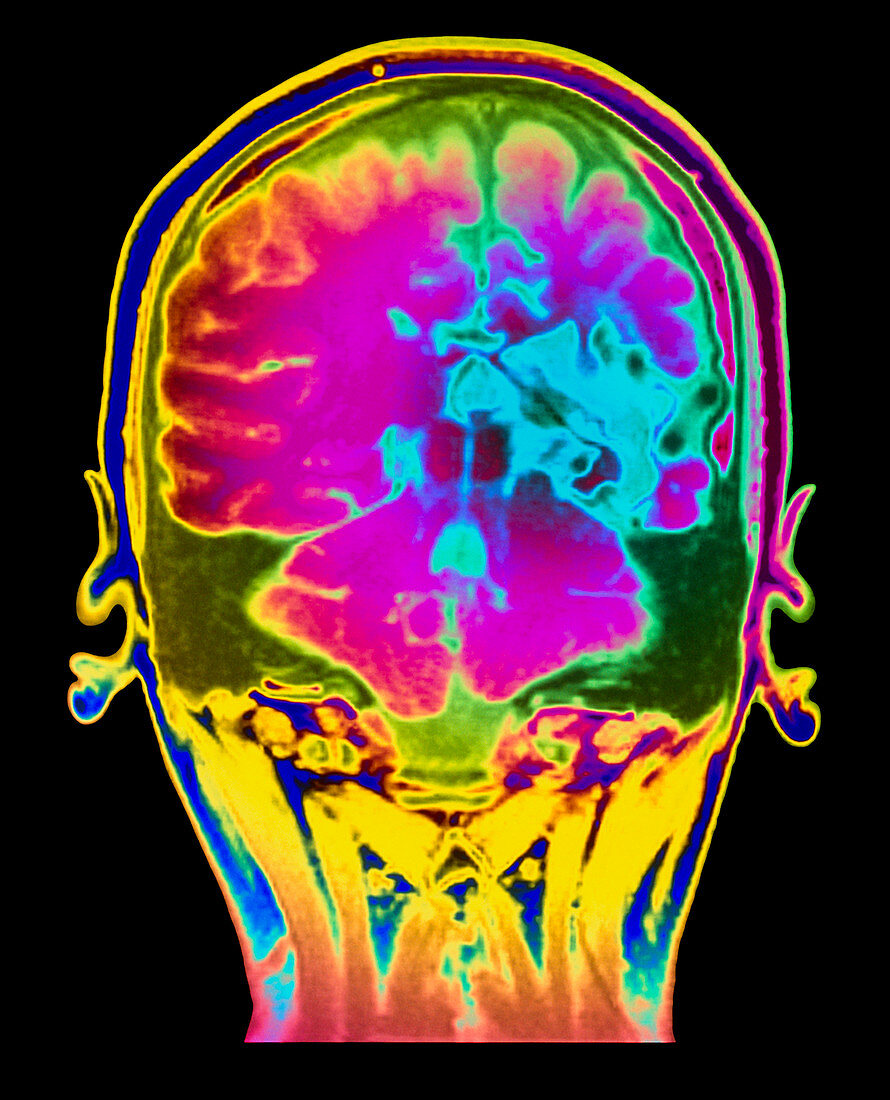
| Brain arteriovenous malfunction. Coloured Magnetic Resonance Imaging (MRI) angiogram of a coronal section through the back of a brain showing arteriovenous malfunction (AVM). The patient,an 18 year old female,suffered this congenital defect from birth. The affected region is at centre right. The blue area shows nerve tissue infarction (death) due to no blood supply. Abnormal blood vessels appear black and circular. Normal brain tissue is pink and orange. AVM may be present at birth or can result from injury or disease. Brain function may be seriously impaired,leading to speech defects or paralysis | |
| Licence : | Droits gérés |
| Crédit: | Science Photo Library / Fraser, Simon / Newcastle Upon Tyne / Royal Victoria Infirmary |
| Taille de l’image : | 4016 px × 4961 px |
| Model Release : | Non requis |
| Property Release : | Non requis |
| Restrictions : | - |
Prix pour cette image À partir de 45 €
Produit vendu
(Calendrier, Carte postale, Carte de vœux, Impression sur textile, Packaging etc)
À partir de 45 €
Usage commercial
(Affichage, Annonce presse, Annonce TV, Carte, Digital - hors rés. sociaux, Digital - rés. sociaux etc)
À partir de 45 €
Éditorial
(Digital, Journal, Livre, Livre pratique, Magazine, Télévision etc)
À partir de 60 €
Usage non-commercial
(Digital - hors rés. sociaux, Digital - rés. sociaux etc)
À partir de 120 €
Mots clés
- angiographie,
- artérioveineux,
- cerveau,
- désordre,
- état,
- I.R.M.,
- imagerie par résonnance magnétique,
- infarctus,
- IRM,
- maladie,
- malformation artérioveineuse,
- médecine,
- médical,
- médicale,
- neuro-imagerie,
- neuroimagerie,
- soins de santé,
- trouble,
- trouble cérébral,
- trouble cerveau,
- vaisseau sanguin,
- vasculaire