Colour PET scans of the brain of a stroke patient
Numéro d’image : 11838241
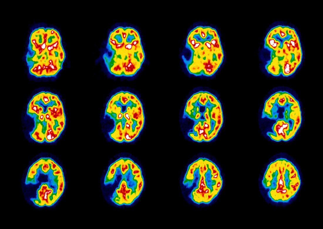
| Stroke. Coloured Positron Emission Tomography (PET) scans of the brain of a stroke patient. Twelve horizontal slices are shown,from deep brain (top left) to superficial (lower right). A lesion is seen in the left hemisphere (dark blue),an area of brain damage with reduced blood flow due to stroke. Colour-coding is: high brain activity (white); low activity (blue). Stroke,or cerebrovascular accident (CVA),is a rupture or blockage of a blood vessel causing brain damage such as loss of speech or paralysis. PET scanning uses an injected radioactive tracer (H2.O15) to show variation in metabolic activity in the brain | |
| Licence : | Droits gérés |
| Crédit: | Science Photo Library / Wellcome Dept. of Cognitive Neurology |
| Taille de l’image : | 3543 px × 2516 px |
| Model Release : | Non requis |
| Property Release : | Non requis |
| Restrictions : | - |
Prix pour cette image À partir de 45 €
Produit vendu
(Calendrier, Carte postale, Carte de vœux, Impression sur textile, Packaging etc)
À partir de 45 €
Usage commercial
(Affichage, Annonce presse, Annonce TV, Carte, Digital - hors rés. sociaux, Digital - rés. sociaux etc)
À partir de 45 €
Éditorial
(Digital, Journal, Livre, Livre pratique, Magazine, Télévision etc)
À partir de 60 €
Usage non-commercial
(Digital - hors rés. sociaux, Digital - rés. sociaux etc)
À partir de 120 €
Mots clés
- A.V.C,
- A.V.C.,
- accident cérébrovasculaire,
- accident vasculaire cérébral,
- animal familier,
- attaque cérébrale,
- AVC,
- cerveau,
- désordre,
- état,
- maladie,
- médecine,
- médical,
- médicale,
- neuro-imagerie,
- neuroimagerie,
- odyssée,
- pet scan,
- soins de santé,
- T.E.P.,
- TEP,
- tomographie par émission de positrons,
- trouble,
- vaisseau sanguin,
- vasculaire