X-ray tomography scan of brain showing haemorrhage
Numéro d’image : 11838220
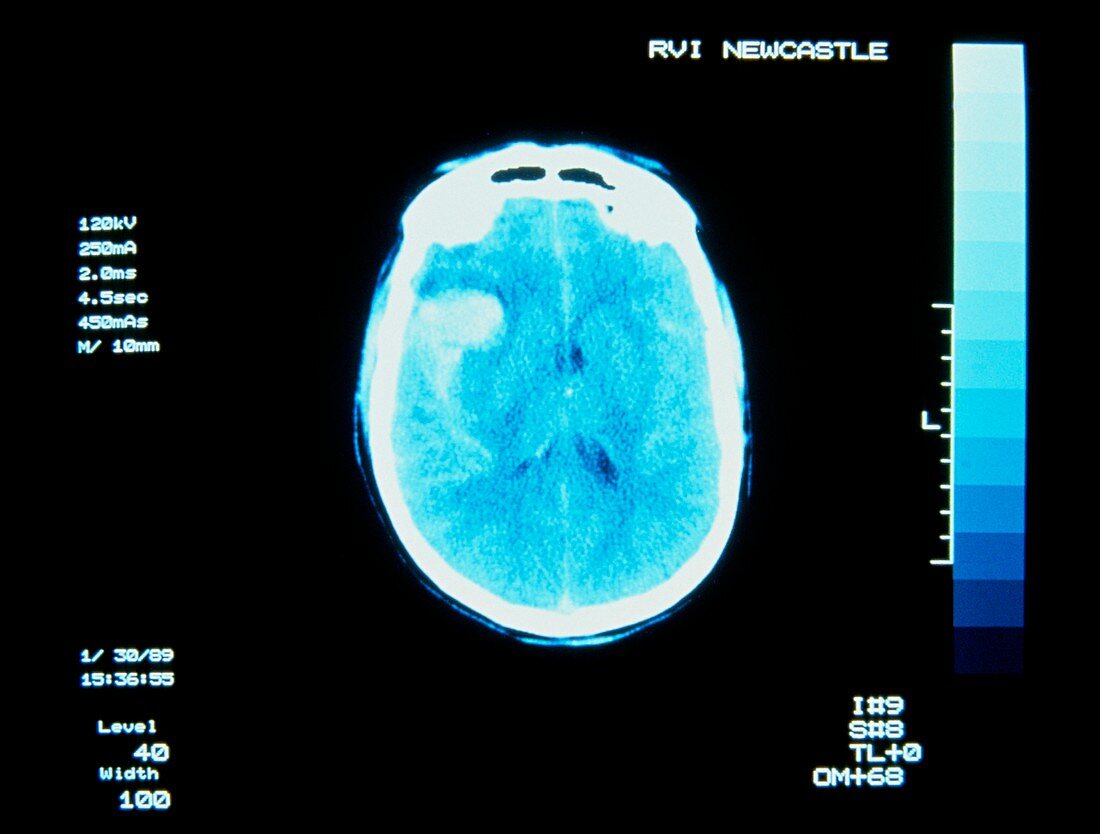
| Computed X-ray tomography (CT) scan of the brain showing a subarachnoid haemorrhage,the result of a ruptured blood vessel & one cause of stroke. The haemorrhage appears as the light area over the right side of the brain (left on image). In CT,a narrow X-ray beam is directed through the subject towards a diametrically-opposed detector. A series of "slices" is made,with source & detector moving synchronously around the subject. Measurements of transmitted X-rays are processed by computer to reveal how elements of tissue in each "slice" attenuate X-rays - the basis for constructing the image. Courtesy of Radiography Dept.,Royal Victoria Infirmary,Newcastle-upon-Tyne | |
| Licence : | Droits gérés |
| Crédit: | Science Photo Library / Fraser, Simon |
| Taille de l’image : | 4843 px × 3670 px |
| Model Release : | Non requis |
| Property Release : | Non requis |
| Restrictions : | - |
Prix pour cette image À partir de 45 €
Produit vendu
(Calendrier, Carte postale, Carte de vœux, Impression sur textile, Packaging etc)
À partir de 45 €
Usage commercial
(Affichage, Annonce presse, Annonce TV, Carte, Digital - hors rés. sociaux, Digital - rés. sociaux etc)
À partir de 45 €
Éditorial
(Digital, Journal, Livre, Livre pratique, Magazine, Télévision etc)
À partir de 60 €
Usage non-commercial
(Digital - hors rés. sociaux, Digital - rés. sociaux etc)
À partir de 120 €