Computed tomography scan of lung cancer biopsy
Numéro d’image : 11837838
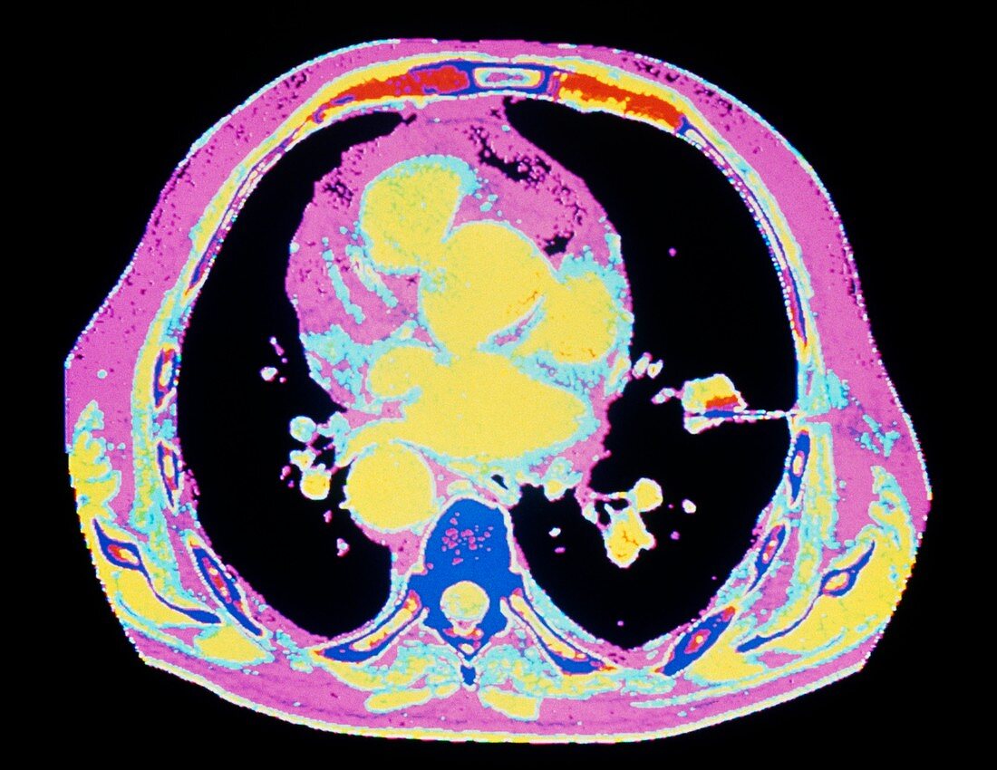
| Lung cancer biopsy. Coloured computed tomography scan of a patient's chest showing a biopsy needle (blue,centre right) taking a tissue sample from a tumour (yellow and red) in the lung. The scan shows a horizontal slice through the chest. Black areas are normal lung tissue. The yellow area in the centre is the heart and its blood vessels. The spine appears in the lower centre (dark blue). Biopsies are carried out to obtain tissue samples for microscope examination. This makes it possible to establish whether a tumour is benign or,as in this case,malignant. Lung cancer is one of the most common and dangerous forms of cancer. The main single cause is cigarette smoking | |
| Licence : | Droits gérés |
| Crédit: | Science Photo Library / CNRI |
| Taille de l’image : | 4785 px × 3697 px |
| Model Release : | Non requis |
| Property Release : | Non requis |
| Restrictions : | - |
Prix pour cette image À partir de 45 €
Produit vendu
(Calendrier, Carte postale, Carte de vœux, Impression sur textile, Packaging etc)
À partir de 45 €
Usage commercial
(Affichage, Annonce presse, Annonce TV, Carte, Digital - hors rés. sociaux, Digital - rés. sociaux etc)
À partir de 45 €
Éditorial
(Digital, Journal, Livre, Livre pratique, Magazine, Télévision etc)
À partir de 60 €
Usage non-commercial
(Digital - hors rés. sociaux, Digital - rés. sociaux etc)
À partir de 120 €