Coloured MRI scan of a metastatic brain tumour
Numéro d’image : 11837821
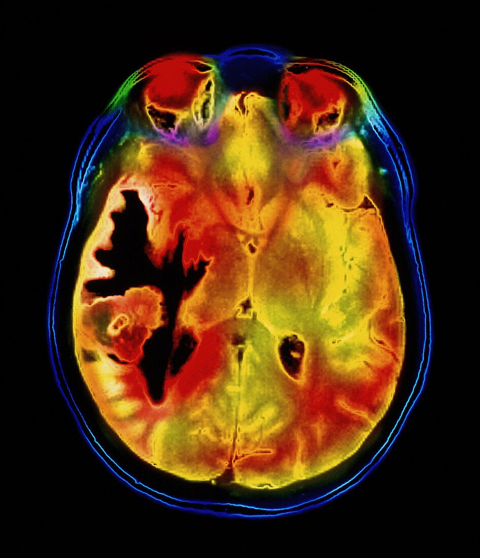
| Brain tumour. Coloured Magnetic Resonance Imaging (MRI) scan of an axial section through the brain of a 42 year old woman,showing a metastatic tumour. At centre left is the tumour (red & yellow) surrounded by damaged fluid-filled tissue (black). This tumour occurs within one cerebral hemisphere; the other hemisphere is at right. The eyeballs (red) are at top. Metastatic cancer is a secondary disease spread from cancer elsewhere in the body. The patient had primary cancer of the colon. Metastatic brain tumours are malignant. Typically they cause brain compression and nerve damage. Surgical removal may be possible | |
| Licence : | Droits gérés |
| Crédit: | Science Photo Library / Fraser, Simon / Newcastle Upon Tyne / Royal Victoria Infirmary |
| Taille de l’image : | 3497 px × 4067 px |
| Model Release : | Non requis |
| Property Release : | Non requis |
| Restrictions : | - |
Prix pour cette image À partir de 45 €
Produit vendu
(Calendrier, Carte postale, Carte de vœux, Impression sur textile, Packaging etc)
À partir de 45 €
Usage commercial
(Affichage, Annonce presse, Annonce TV, Carte, Digital - hors rés. sociaux, Digital - rés. sociaux etc)
À partir de 45 €
Éditorial
(Digital, Journal, Livre, Livre pratique, Magazine, Télévision etc)
À partir de 60 €
Usage non-commercial
(Digital - hors rés. sociaux, Digital - rés. sociaux etc)
À partir de 120 €
Mots clés
- cancer,
- cancer métastatique,
- cancéreux,
- cerveau,
- désordre,
- état,
- I.R.M.,
- imagerie du cancer,
- imagerie par résonnance magnétique,
- IRM,
- maladie,
- malignité,
- malin,
- médecine,
- médical,
- médicale,
- neuro-imagerie,
- neuroimagerie,
- scanner du cerveau,
- soins de santé,
- trouble,
- tumeur,
- tumeur intra-crânienne,
- tumeur intracrânienne,
- tumeur maligne,
- tumeurs cérébrales