CT scan showing kidney cancer
Numéro d’image : 11837803
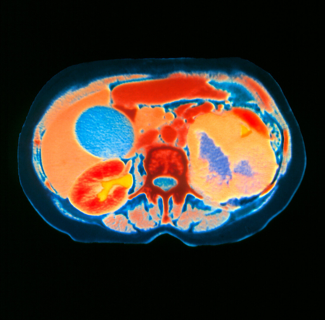
| Kidney cancer. Coloured computed Tomography (CT) scan,axial section,of the human abdomen showing kidney cancer. At centre is a vertebra (rust). The diseased kidney (right on image,orange) is distorted and enlarged by a cancer tumour. This diseased kidney with a blue coloured interior is non-functioning except for a small area excreting into the collecting system (yellow triangle,upper right). At lower left is a normal kidney (red,bean-shaped),that is excreting an X-ray contrast agent (yellow). The liver (brown,left) is next to the normal kidney,with the circular gall bladder (blue) covering the liver | |
| Licence : | Droits gérés |
| Crédit: | Science Photo Library / DEPT. OF CLINICAL RADIOLOGY, SALISBURY DISTRICT HOSPITAL |
| Taille de l’image : | 3238 px × 3189 px |
| Model Release : | Non requis |
| Property Release : | Non requis |
| Restrictions : | - |
Prix pour cette image À partir de 45 €
Produit vendu
(Calendrier, Carte postale, Carte de vœux, Impression sur textile, Packaging etc)
À partir de 45 €
Usage commercial
(Affichage, Annonce presse, Annonce TV, Carte, Digital - hors rés. sociaux, Digital - rés. sociaux etc)
À partir de 45 €
Éditorial
(Digital, Journal, Livre, Livre pratique, Magazine, Télévision etc)
À partir de 60 €
Usage non-commercial
(Digital - hors rés. sociaux, Digital - rés. sociaux etc)
À partir de 120 €