MRI image of an acoustic neuroma tumour
Numéro d’image : 11837746
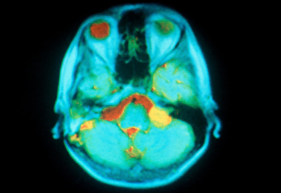
| Nuclear magnetic resonance (NMR) image of the brain of person suffering from an acoustic neuroma,a tumour which occurs between the inner ear & the brain,growing from the fibrous coverings of a peripheral nerve. In this image the growth appears as the yellow region on the right side of the brain. Also visible are the eyes (top); the vitreous humour in the eyeballs appears red in the left eye,yellow in the right. The colour coding represents a parameter in NMR imaging known as T1 (relaxation time),with blue a short T1 & red a larger T1 value | |
| Licence : | Droits gérés |
| Crédit: | Science Photo Library / HAMMERSMITH HOSPITAL MEDICAL SCHOOL / GEC RESEARCH |
| Taille de l’image : | 4615 px × 3176 px |
| Model Release : | Non requis |
| Property Release : | Non requis |
| Restrictions : | - |
Prix pour cette image À partir de 45 €
Produit vendu
(Calendrier, Carte postale, Carte de vœux, Impression sur textile, Packaging etc)
À partir de 45 €
Usage commercial
(Affichage, Annonce presse, Annonce TV, Carte, Digital - hors rés. sociaux, Digital - rés. sociaux etc)
À partir de 45 €
Éditorial
(Digital, Journal, Livre, Livre pratique, Magazine, Télévision etc)
À partir de 60 €
Usage non-commercial
(Digital - hors rés. sociaux, Digital - rés. sociaux etc)
À partir de 120 €