Cancer cell division
Numéro d’image : 11837464
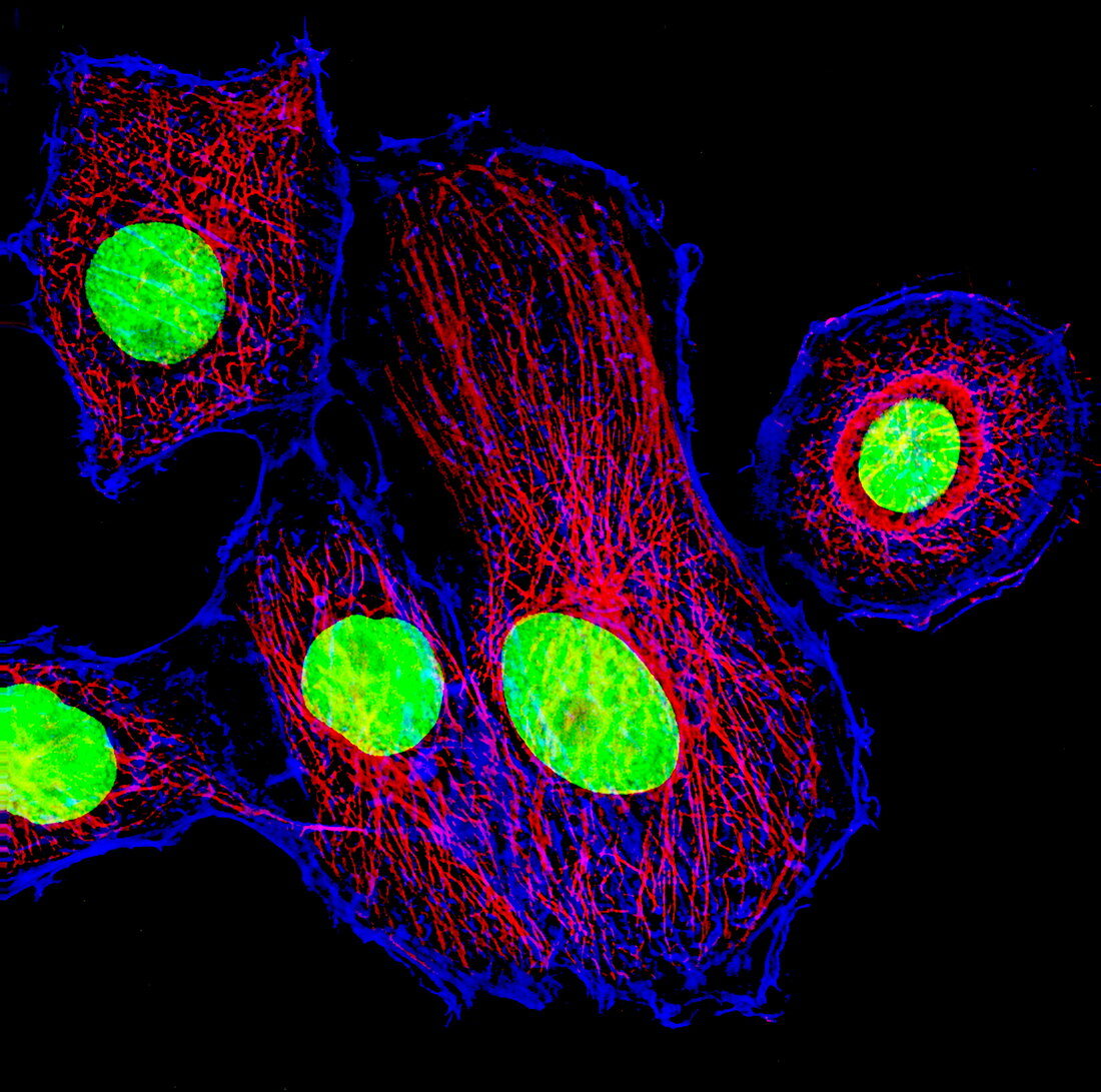
| Cancer cell division. Immunofluorescent light micrograph of cancer cells during the interphase stage of cell division (mitosis). Interphase is the resting stage of mitosis,when the cell's deoxyribonucleic acid (DNA,green) is replicated. Also seen is the actin cytoskeleton (red) and microtubules (red). Proteins are also synthesised and the cell increases in size. During mitosis a cell produces two genetically- identical daughter cells. Immunofluorescence uses antibodies to attach fluorescent dyes to specific cell tissues | |
| Licence : | Droits gérés |
| Crédit: | Science Photo Library / DR PAUL ANDREWS, UNIVERSITY OF DUNDEE |
| Taille de l’image : | 2967 px × 2942 px |
| Model Release : | Non requis |
| Property Release : | Non requis |
| Restrictions : | - |
Prix pour cette image À partir de 45 €
Produit vendu
(Calendrier, Carte postale, Carte de vœux, Impression sur textile, Packaging etc)
À partir de 45 €
Usage commercial
(Affichage, Annonce presse, Annonce TV, Carte, Digital - hors rés. sociaux, Digital - rés. sociaux etc)
À partir de 45 €
Éditorial
(Digital, Journal, Livre, Livre pratique, Magazine, Télévision etc)
À partir de 60 €
Usage non-commercial
(Digital - hors rés. sociaux, Digital - rés. sociaux etc)
À partir de 120 €
Mots clés
- A.D.N.,
- acide désoxyribonucléique,
- ADN,
- anormal,
- biologie,
- biologique,
- cancer,
- cancéreux,
- cellule,
- cellules,
- cinq,
- cycle,
- cyclique,
- cytologie,
- cytologique,
- division cellulaire,
- étape,
- génétique,
- immunofluorescence,
- interphase,
- malsain,
- microscope optique,
- microscopie optique,
- microtubules,
- mitose,
- oncologie,
- oncologique,
- phase,
- quintette,
- répétitif,
- répliquer,
- repos,
- se reposer,
- stade