Immunofluorecent LM of human colon cancer cells
Numéro d’image : 11837229
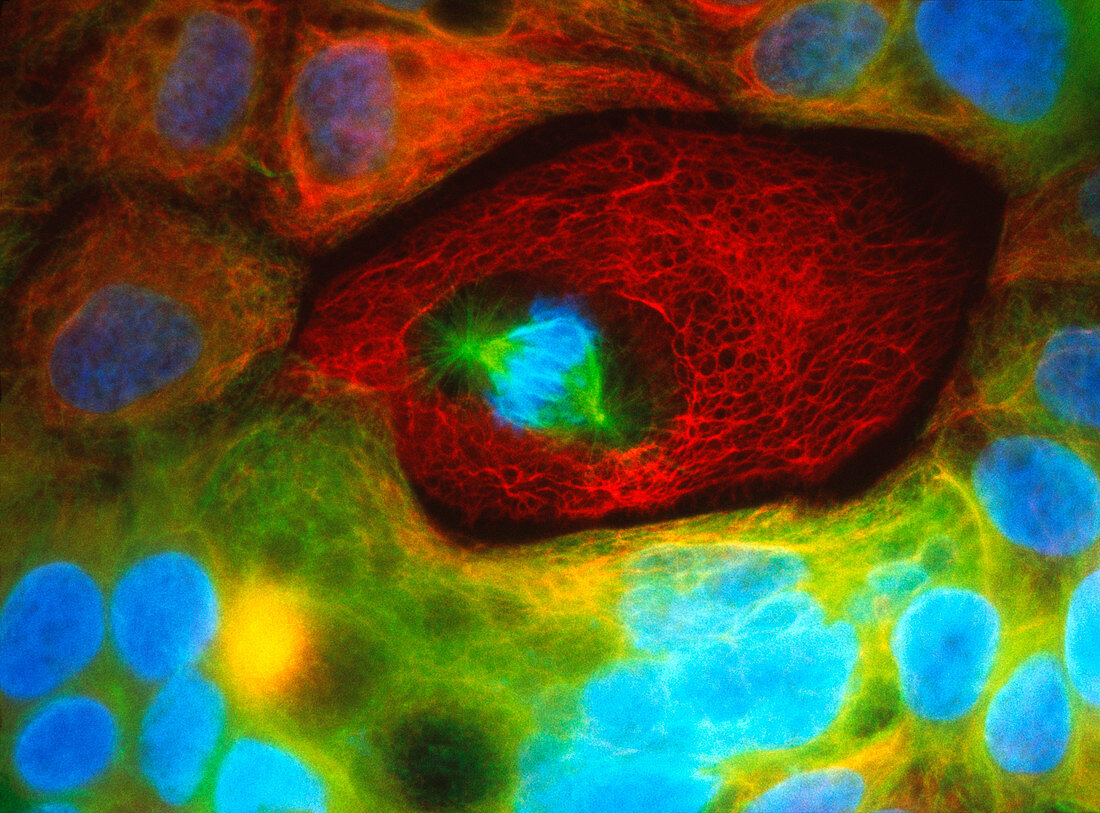
| Colon cancer. Immunofluorescent light micrograph of human colon cancer cells. The nucleus of each cell is blue,surrounded by a fibrous mass of protein. Tubulin (green) is a protein used by cells during cell division,myosin (red) is muscle fibre. Mitotic cell division (at centre) is occurring. Blue condensed chromosomes are being "split" apart by two tubulin spindles. Such irregular-shaped cells with large nuclei and cell divisions are characteristic of cancer cells. Colonic cancer is a common form of cancer. Immuno- fluorescence is a technique that uses antibodies to attach fluorescent dyes to specific parts of a cell. Magnification: x374 at 4.5x6cm size | |
| Licence : | Droits gérés |
| Crédit: | Science Photo Library / Kedersha, Nancy |
| Taille de l’image : | 4370 px × 3230 px |
| Model Release : | Non requis |
| Property Release : | Non requis |
| Restrictions : | - |
Prix pour cette image À partir de 45 €
Produit vendu
(Calendrier, Carte postale, Carte de vœux, Impression sur textile, Packaging etc)
À partir de 45 €
Usage commercial
(Affichage, Annonce presse, Annonce TV, Carte, Digital - hors rés. sociaux, Digital - rés. sociaux etc)
À partir de 45 €
Éditorial
(Digital, Journal, Livre, Livre pratique, Magazine, Télévision etc)
À partir de 60 €
Usage non-commercial
(Digital - hors rés. sociaux, Digital - rés. sociaux etc)
À partir de 120 €
Mots clés
- agrandissement,
- cancer,
- cancer du côlon,
- cancéreux,
- cellule cancéreuse,
- cellules cancéreuses,
- colon,
- désordre,
- du côlon,
- état,
- images,
- immunofluorescence,
- maladie,
- malignité,
- malin,
- médecine,
- médical,
- médicale,
- microscope optique,
- microscopie optique,
- mitose,
- myosine,
- photos au microscope,
- soins de santé,
- sujets,
- trouble,
- tubuline,
- tumeur maligne