Light micrograph of secondary bone cancer
Numéro d’image : 11837208
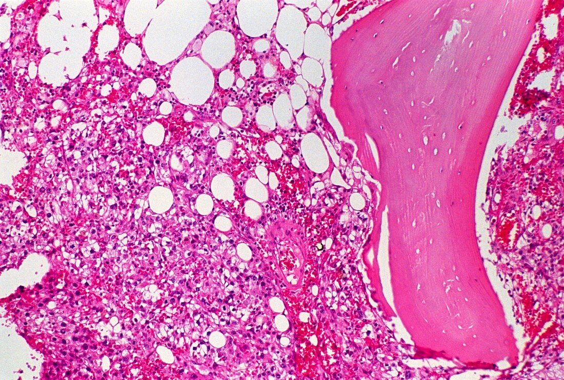
| Secondary bone cancer. Light micrograph of a secondary (metastatic) cancer tumour developing in bone. This malignant tumour,called a carcinoma,has grown from cancer cells that have spread from the kidney. Cancer cells here are pink with large nuclei,typical of rapidly dividing cells. On the right is a piece of bone (pink). Clear spaces are fat cells. This type of tumour contains numerous blood vessels,which here are filled with red blood cells. The tumour has replaced any normal bone marrow. Sample stained with hematoxylin and eosin. Magnification x125 at 35mm size | |
| Licence : | Droits gérés |
| Crédit: | Science Photo Library / Walker, Dr. E. |
| Taille de l’image : | 5135 px × 3461 px |
| Model Release : | Non requis |
| Property Release : | Non requis |
| Restrictions : | - |
Prix pour cette image À partir de 45 €
Produit vendu
(Calendrier, Carte postale, Carte de vœux, Impression sur textile, Packaging etc)
À partir de 45 €
Usage commercial
(Affichage, Annonce presse, Annonce TV, Carte, Digital - hors rés. sociaux, Digital - rés. sociaux etc)
À partir de 45 €
Éditorial
(Digital, Journal, Livre, Livre pratique, Magazine, Télévision etc)
À partir de 60 €
Usage non-commercial
(Digital - hors rés. sociaux, Digital - rés. sociaux etc)
À partir de 120 €