LM of secondary bone cancer (carcinoma)
Numéro d’image : 11837204
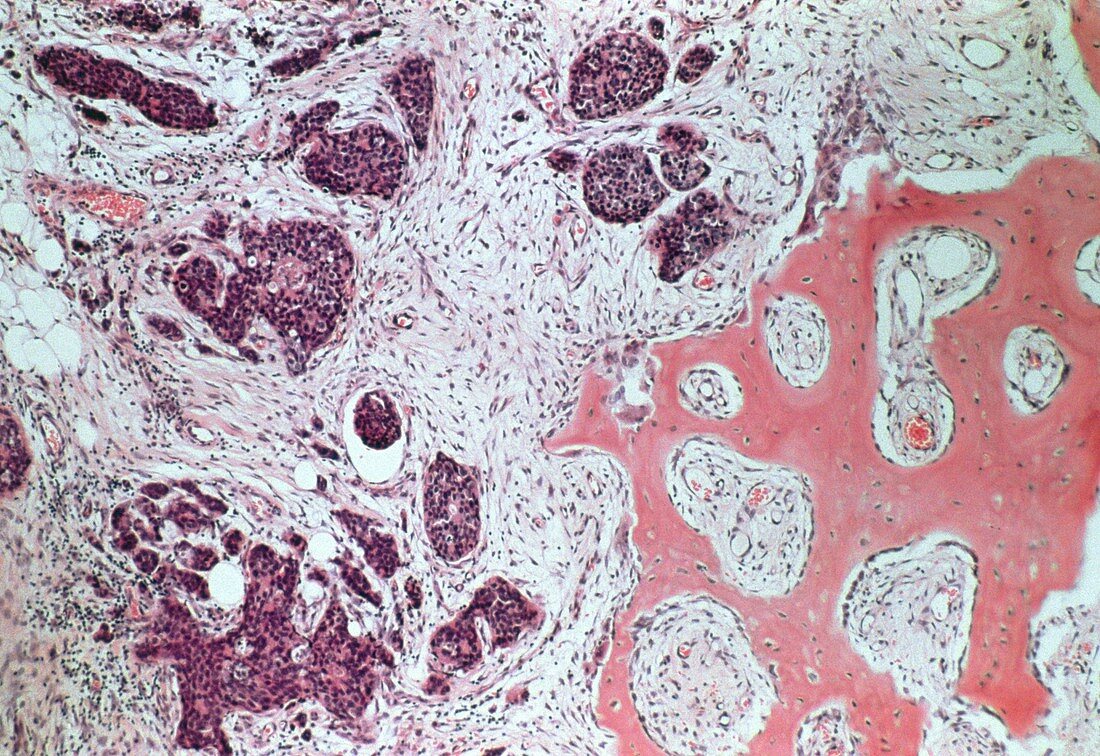
| Secondary bone cancer. Light micrograph of a secondary (metastatic) cancer tumour developing in bone. This malignant tumour,called a carcinoma,has grown from cancer cells that have spread from the breast. Cancerous cells are dark purple,with large nuclei typical of rapidly dividing cells. On the right is a fragment of bone (pink). The inter- vening pale cells are connective tissue laid down in response to the tumour. White blood cells (dark dots on left) have infiltrated as a result of in- flammation. There is no residual bone marrow. Breast cancer is the most common cancer in women. Sample stained with hematoxylin and eosin. Magnification x125 at 35mm size | |
| Licence : | Droits gérés |
| Crédit: | Science Photo Library / Walker, Dr. E. |
| Taille de l’image : | 5091 px × 3500 px |
| Model Release : | Non requis |
| Property Release : | Non requis |
| Restrictions : | - |
Prix pour cette image À partir de 45 €
Produit vendu
(Calendrier, Carte postale, Carte de vœux, Impression sur textile, Packaging etc)
À partir de 45 €
Usage commercial
(Affichage, Annonce presse, Annonce TV, Carte, Digital - hors rés. sociaux, Digital - rés. sociaux etc)
À partir de 45 €
Éditorial
(Digital, Journal, Livre, Livre pratique, Magazine, Télévision etc)
À partir de 60 €
Usage non-commercial
(Digital - hors rés. sociaux, Digital - rés. sociaux etc)
À partir de 120 €