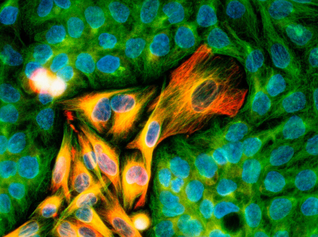
Immunofluorescent LM of melanoma cancer cells
Numéro d’image : 11837155

| Immunofluorescent Light Micrograph of melanoma cancer cells invading the skin epithelium,seen in a culture preparation. At centre is a tumour of cancer (orange) derived from melanin-forming cells of the skin. Normal epithelial skin cells are green. Each cell nucleus stains blue. Melanoma is a highly malignant cancer consisting of large undifferentiated cells with a capacity to divide rapidly and invade surrounding healthy tissue. Immunofluorescence is a staining technique which uses antibodies to attach fluorescent dyes to specific tissues and to molecules within the cell. Magnification: x400 at 35mm size,x750 at 6x4.5cm | |
| Licence : | Droits gérés |
| Crédit: | Science Photo Library / Kedersha, Nancy |
| Taille de l’image : | 4240 px × 3173 px |
| Model Release : | Non requis |
| Property Release : | Non requis |
| Restrictions : | - |
Prix pour cette image À partir de 45 €
Produit vendu
(Calendrier, Carte postale, Carte de vœux, Impression sur textile, Packaging etc)
À partir de 45 €
Usage commercial
(Affichage, Annonce presse, Annonce TV, Carte, Digital - hors rés. sociaux, Digital - rés. sociaux etc)
À partir de 45 €
Éditorial
(Digital, Journal, Livre, Livre pratique, Magazine, Télévision etc)
À partir de 60 €
Usage non-commercial
(Digital - hors rés. sociaux, Digital - rés. sociaux etc)
À partir de 120 €
Mots clés
- agrandissement,
- cancer,
- cancer de la peau,
- cancéreux,
- cellule,
- cellule cancéreuse,
- désordre,
- état,
- images,
- immunofluorescence,
- maladie,
- malignité,
- malin,
- médecine,
- médical,
- médicale,
- melanoma,
- mélanome,
- microscope optique,
- microscopie optique,
- photos au microscope,
- soins de santé,
- sujets,
- trouble,
- tumeur maligne
