False-col SEM of T-lymphocyte killer cell
Numéro d’image : 11837096
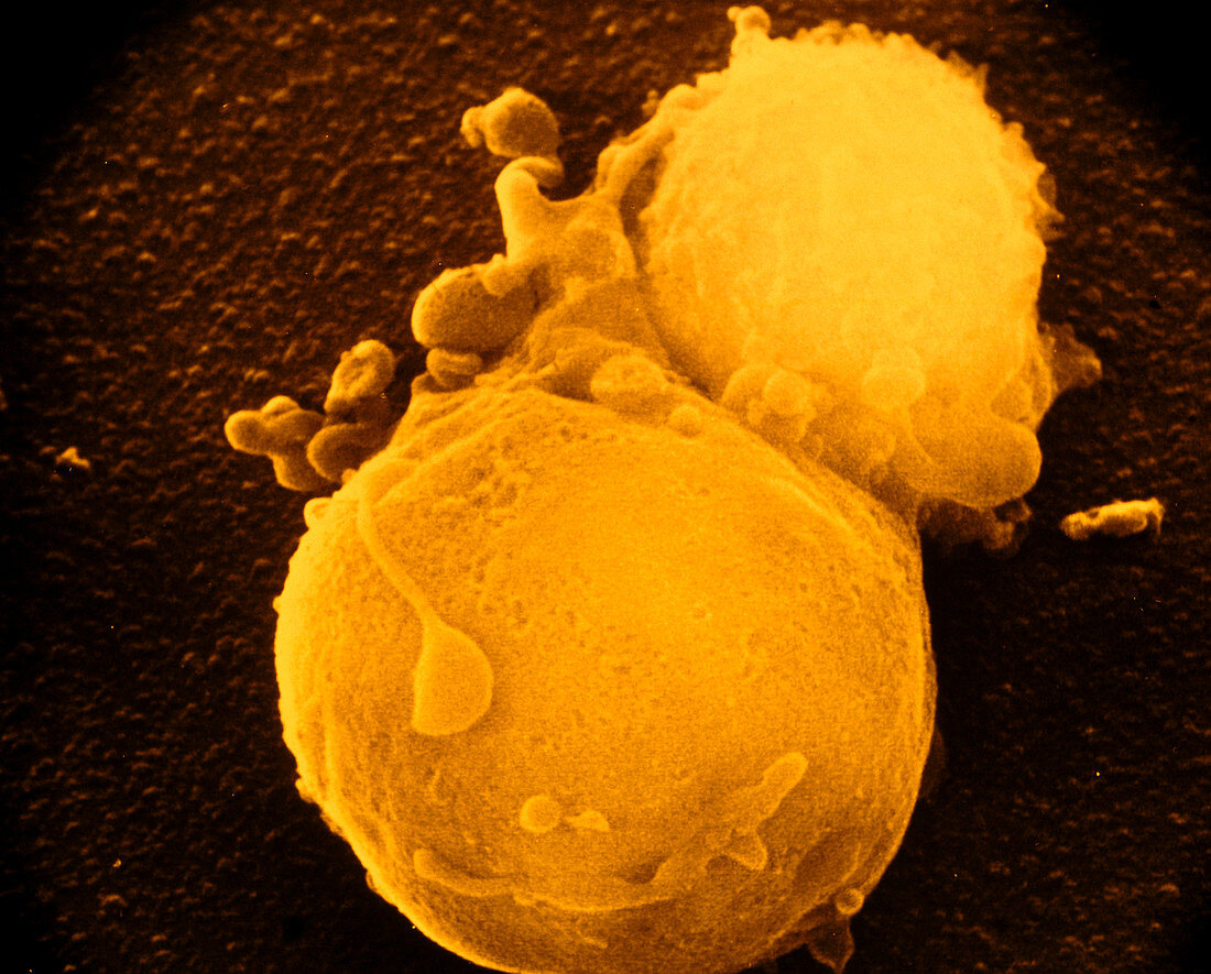
| False-colour scanning electron micrograph of a small T-lymphocyte killer cell (top) which has successfully attacked a large tumour or cancer cell (bottom). The surface of the tumour cell has lost its normal covering of small projections called microvilli,which is an indication that it is losing the battle with the lymphocyte. After it is attacked by the lymphocyte,a white blood cell,the tumour cell defends itself by producing blisters,or blebs (some present here),to prevent the lymphocyte making intimate contact. If this strategy is not successful,the lymphocyte may kill the tumour cell,as here. Magnification: X 2,500 at 35mm size | |
| Licence : | Droits gérés |
| Crédit: | Science Photo Library / Liepins, Dr. Andrejs |
| Taille de l’image : | 4606 px × 3707 px |
| Model Release : | Non requis |
| Property Release : | Non requis |
| Restrictions : | - |
Prix pour cette image À partir de 45 €
Produit vendu
(Calendrier, Carte postale, Carte de vœux, Impression sur textile, Packaging etc)
À partir de 45 €
Usage commercial
(Affichage, Annonce presse, Annonce TV, Carte, Digital - hors rés. sociaux, Digital - rés. sociaux etc)
À partir de 45 €
Éditorial
(Digital, Journal, Livre, Livre pratique, Magazine, Télévision etc)
À partir de 60 €
Usage non-commercial
(Digital - hors rés. sociaux, Digital - rés. sociaux etc)
À partir de 120 €