SEM of T-lymphocyte killer cell
Numéro d’image : 11837093
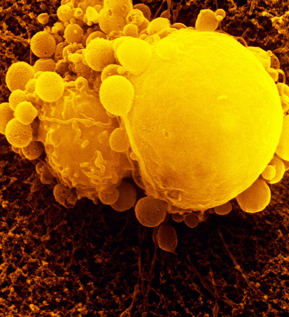
| False-colour scannning electron micrograph of a T-lymphocyte killer cell (small cell on left),attacking a large cancer tumour cell. The T-ymphocyte,a white blood cell,must make intimate contact with the tumour cell. It does so by recognizing antigens on the tumour's surface. Following contact the tumour cell undergoes distinct structural changes; loss of microvilli (small projections,lost here),perforation and ultimately death. The tumour cell may survive,however,by budding off a number of blebs or blisters,which form a protective barrier between itself & the lymphocyte,preventing further contact. Magnification: X 2,500 at 35mm size | |
| Licence : | Droits gérés |
| Crédit: | Science Photo Library / Liepins, Dr. Andrejs |
| Taille de l’image : | 2972 px × 3259 px |
| Model Release : | Non requis |
| Property Release : | Non requis |
| Restrictions : | - |
Prix pour cette image À partir de 45 €
Produit vendu
(Calendrier, Carte postale, Carte de vœux, Impression sur textile, Packaging etc)
À partir de 45 €
Usage commercial
(Affichage, Annonce presse, Annonce TV, Carte, Digital - hors rés. sociaux, Digital - rés. sociaux etc)
À partir de 45 €
Éditorial
(Digital, Journal, Livre, Livre pratique, Magazine, Télévision etc)
À partir de 60 €
Usage non-commercial
(Digital - hors rés. sociaux, Digital - rés. sociaux etc)
À partir de 120 €
Mots clés
- attaquer,
- cancéreux,
- cellule cancéreuse,
- cellule t,
- cellule tumorale,
- désordre,
- état,
- globule blanc,
- lymphocyte,
- lymphocyte t,
- lymphocyte-t,
- M.E.B.,
- maladie,
- malignité,
- malin,
- MEB,
- médecine,
- médical,
- médicale,
- microscope électronique à balayage,
- phagocytose,
- phagocytosis,
- soins de santé,
- trouble,
- tumeur maligne