Brain cyst in a young child
Numéro d’image : 11836241
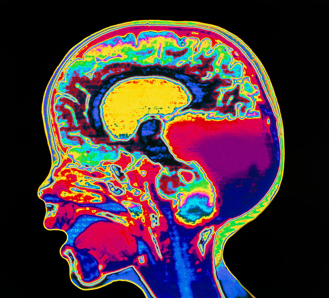
| Brain cyst. Coloured magnetic resonance imaging (MRI) scan of a section through the brain of an 11 month old child. At lower right a cyst (red) has formed in the region of the cerebellum. The brain's ventricle (yellow) can also be seen in the image. Cysts are abnormal lumps or swellings filled with fluid or semi-solid material. They can occur in any organ or tissue. Cysts are usually harmless,but if they grow disproportionately large they may disrupt the function of the tissue. MRI scanners produce slice images though the body. The images are built up from radio signals and a magnetic field | |
| Licence : | Droits gérés |
| Crédit: | Science Photo Library / Kulyk, Mehau |
| Taille de l’image : | 4724 px × 4275 px |
| Model Release : | Non requis |
| Property Release : | Non requis |
| Restrictions : | - |
Prix pour cette image À partir de 45 €
Produit vendu
(Calendrier, Carte postale, Carte de vœux, Impression sur textile, Packaging etc)
À partir de 45 €
Usage commercial
(Affichage, Annonce presse, Annonce TV, Carte, Digital - hors rés. sociaux, Digital - rés. sociaux etc)
À partir de 45 €
Éditorial
(Digital, Journal, Livre, Livre pratique, Magazine, Télévision etc)
À partir de 60 €
Usage non-commercial
(Digital - hors rés. sociaux, Digital - rés. sociaux etc)
À partir de 120 €
Mots clés
- cerebrum,
- cerveau,
- coupé,
- coupe-circuit,
- découpé,
- découpes,
- désordre,
- détourages,
- détouré,
- disjoncteur,
- enfant,
- état,
- I.R.M.,
- imagerie par résonnance magnétique,
- IRM,
- kyste,
- kyste au cerveau,
- kyste cérébral,
- maladie,
- médecine,
- médical,
- médicale,
- neuro-imagerie,
- neuroimagerie,
- silhouette,
- soins de santé,
- trouble,
- ventricule