Coloured 3-D MRI scan of a brain cyst
Numéro d’image : 11836227
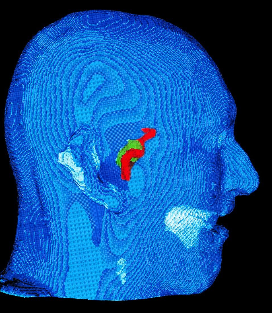
| Brain cyst. Coloured three-dimensional magnetic resonance imaging (MRI) scan of a brain cyst as seen through a MAGI virtual reality microscope. The cyst is green and part of an internal carotid artery is red. The MRI scan of the cyst is seen on a coloured three-dimensional computed tomography (CT) head scan. This is a simulated view of the MAGI (microscope-assisted guided interventions) virtual reality system. With MAGI,surgeons see a superimposed image of previously scanned struc- tures beneath the skin while looking through a microscope during surgery. This helps the surgeon to find the cyst while avoiding the artery. MAGI was developed by teams from London University,UK | |
| Licence : | Droits gérés |
| Crédit: | Science Photo Library / Tompkinson, Geoff |
| Taille de l’image : | 3848 px × 4429 px |
| Model Release : | Non requis |
| Property Release : | Non requis |
| Restrictions : | - |
Prix pour cette image À partir de 45 €
Produit vendu
(Calendrier, Carte postale, Carte de vœux, Impression sur textile, Packaging etc)
À partir de 45 €
Usage commercial
(Affichage, Annonce presse, Annonce TV, Carte, Digital - hors rés. sociaux, Digital - rés. sociaux etc)
À partir de 45 €
Éditorial
(Digital, Journal, Livre, Livre pratique, Magazine, Télévision etc)
À partir de 60 €
Usage non-commercial
(Digital - hors rés. sociaux, Digital - rés. sociaux etc)
À partir de 120 €
Mots clés
- 3 D,
- 3 dimensions,
- 3-D,
- 3D,
- artère carotide,
- cerveau,
- désordre,
- état,
- I.R.M.,
- imagerie par résonnance magnétique,
- IRM,
- kyste,
- kyste au cerveau,
- kyste cérébral,
- maladie,
- médecine,
- médical,
- médicale,
- neuro-imagerie,
- neuroimagerie,
- opération chirurgicale,
- réalité virtuelle,
- soins de santé,
- trois dimensions,
- trouble,
- vaisseau sanguin