SEM of intestine showing coeliac disease
Numéro d’image : 11836134
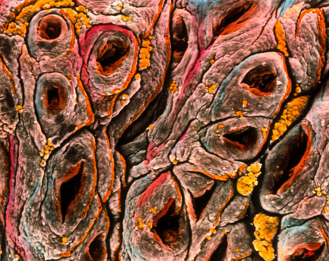
| Coeliac disease. Coloured Scanning Electron Micrograph (SEM) of the wall of the small intestine,showing coeliac disease. The human intestinal wall (mucosa) appears flat and atrophied due to the loss of villi which normally project from the wall. Large holes correspond to the openings of intestinal glands. Yellow particles are migrating leucocytes (white blood cells). Coeliac disease (or gluten enteropathy) is an uncommon genetic condition in which the lining of the intestine reacts to gluten,a protein found in cereals. This causes malabsorption,mineral and vitamin deficiency. Magnification: x350 at 6x7cm size | |
| Licence : | Droits gérés |
| Crédit: | Science Photo Library / PROFESSORS P.M. MOTTA & F.M. MAGLIOCCA |
| Taille de l’image : | 3088 px × 2455 px |
| Model Release : | Non requis |
| Property Release : | Non requis |
| Restrictions : | - |
Prix pour cette image À partir de 45 €
Produit vendu
(Calendrier, Carte postale, Carte de vœux, Impression sur textile, Packaging etc)
À partir de 45 €
Usage commercial
(Affichage, Annonce presse, Annonce TV, Carte, Digital - hors rés. sociaux, Digital - rés. sociaux etc)
À partir de 45 €
Éditorial
(Digital, Journal, Livre, Livre pratique, Magazine, Télévision etc)
À partir de 60 €
Usage non-commercial
(Digital - hors rés. sociaux, Digital - rés. sociaux etc)
À partir de 120 €