Coloured SEM of a liver cell affected by cirrhosis
Numéro d’image : 11836125
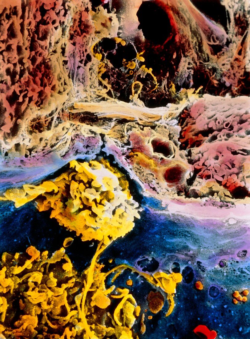
| Cirrhosis. Coloured scanning electron micrograph of a human liver cell (red-brown),also known as hepatocyte,showing abnormal features caused by cirrhosis. It is a disease in which bands of connective tissue (pale orange upper centre) break up the normal structure of the liver severely impairing its functions. The surface of the liver cell (upper centre right) is abnormal showing irregular microvilli and larger evaginations. Two activated macrophages (Kupffer cells,yellow) are seen in a capillary (blue). The wall of the capillary (pale purple) is very thick and fenestrated. Magnification: x2250 at 6x7cm size | |
| Licence : | Droits gérés |
| Crédit: | Science Photo Library / PROFESSORS P. MOTTA, M. NISHI & T. FUJITA |
| Taille de l’image : | 3591 px × 4867 px |
| Model Release : | Non requis |
| Property Release : | Non requis |
| Restrictions : | - |
Prix pour cette image À partir de 45 €
Produit vendu
(Calendrier, Carte postale, Carte de vœux, Impression sur textile, Packaging etc)
À partir de 45 €
Usage commercial
(Affichage, Annonce presse, Annonce TV, Carte, Digital - hors rés. sociaux, Digital - rés. sociaux etc)
À partir de 45 €
Éditorial
(Digital, Journal, Livre, Livre pratique, Magazine, Télévision etc)
À partir de 60 €
Usage non-commercial
(Digital - hors rés. sociaux, Digital - rés. sociaux etc)
À partir de 120 €