X-ray view of knee joint with osteoarthritis
Numéro d’image : 11835098
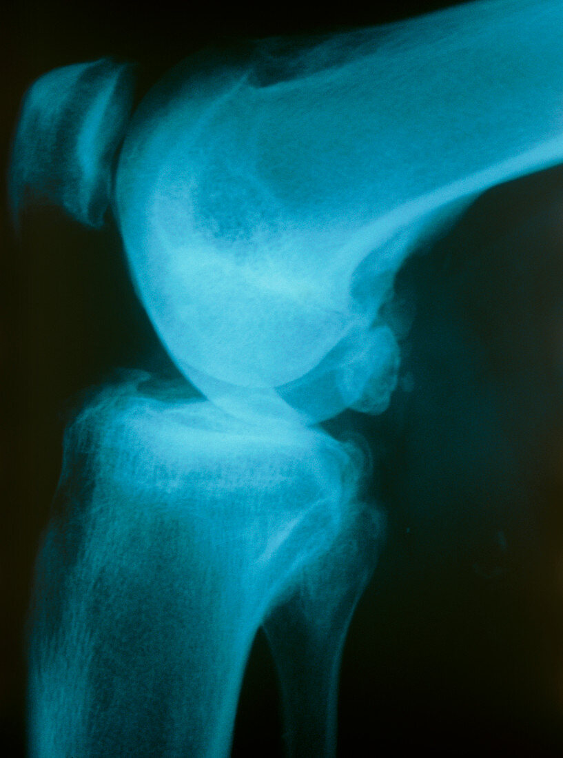
| Osteoarthritis of knee. X-ray image of a side view of the bending knee joint of a 70 year-old woman with osteoarthritis. The lower end of the femur (thigh bone) is seen at top,this joins to the upper end of the tibia (shin bone,lower image). In a normal knee joint,a clear space should be seen between the femur and tibia. Osteoarthritis has lead to the loss of intervening joint surface which consists of cartilage and lubricating fluid. The ends of the bones have become severely eroded. A broken fragment of bone is seen at centre right. Osteoarthritis is the most common type of arthritis due mainly to wear and tear on joints in the elderly | |
| Licence : | Droits gérés |
| Crédit: | Science Photo Library / Marazzi, Dr. P. |
| Taille de l’image : | 3440 px × 4634 px |
| Model Release : | Non requis |
| Property Release : | Non requis |
| Restrictions : | - |
Prix pour cette image À partir de 45 €
Produit vendu
(Calendrier, Carte postale, Carte de vœux, Impression sur textile, Packaging etc)
À partir de 45 €
Usage commercial
(Affichage, Annonce presse, Annonce TV, Carte, Digital - hors rés. sociaux, Digital - rés. sociaux etc)
À partir de 45 €
Éditorial
(Digital, Journal, Livre, Livre pratique, Magazine, Télévision etc)
À partir de 60 €
Usage non-commercial
(Digital - hors rés. sociaux, Digital - rés. sociaux etc)
À partir de 120 €
Mots clés
- affection articulaire,
- affection des articulations,
- arthrite,
- arthritique,
- arthritis,
- arthrose,
- articulation,
- articulation du genou,
- désordre,
- état,
- genou,
- maladie,
- maladie articulaire,
- maladie des articulations,
- médecine,
- médical,
- médicale,
- ostéo-,
- osteoarthritis,
- radiographie,
- rayons X,
- soins de santé,
- trouble