Colour MRI scan of knee joint with osteoarthritis
Numéro d’image : 11835094
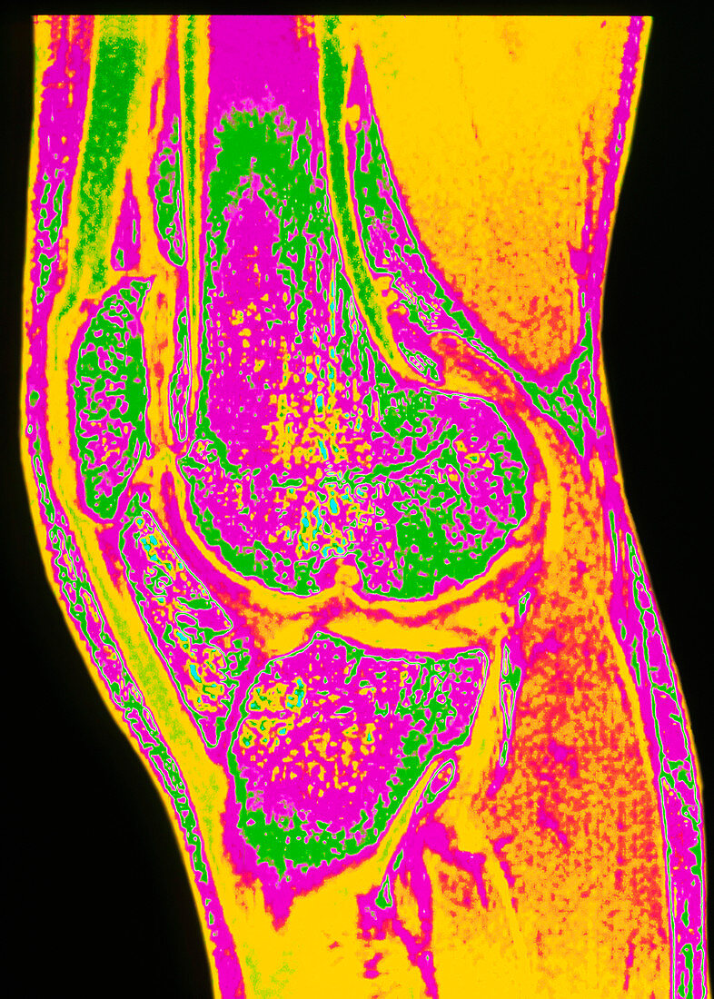
| Osteoarthritis of knee. Colour Magnetic Resonance Imaging (MRI) scan of a side view of the knee joint of a 33 year old male with osteoarthritis. The lower end of the femur (thigh bone) is at centre (pink); it articulates with the upper end of the tibia (shin bone) at lower centre (pink). The patella kneecap is at centre left. Due to osteoarthritis the end of the femur is eroded and jagged; cracks causing weakening in both bones are also visible. Osteoarthritis is the most common type of arthritis due mainly to wear and tear on joints largely affecting the elderly. This patient had haemophilia which is a factor in arthritis | |
| Licence : | Droits gérés |
| Crédit: | Science Photo Library / SIMON FRASER, ROYAL VICTORIA INFIRMARY |
| Taille de l’image : | 2533 px × 3543 px |
| Model Release : | Non requis |
| Property Release : | Non requis |
| Restrictions : | - |
Prix pour cette image À partir de 45 €
Produit vendu
(Calendrier, Carte postale, Carte de vœux, Impression sur textile, Packaging etc)
À partir de 45 €
Usage commercial
(Affichage, Annonce presse, Annonce TV, Carte, Digital - hors rés. sociaux, Digital - rés. sociaux etc)
À partir de 45 €
Éditorial
(Digital, Journal, Livre, Livre pratique, Magazine, Télévision etc)
À partir de 60 €
Usage non-commercial
(Digital - hors rés. sociaux, Digital - rés. sociaux etc)
À partir de 120 €
Mots clés
- affection articulaire,
- affection des articulations,
- arthrite,
- arthritique,
- arthritis,
- arthrose,
- articulation,
- articulation du genou,
- désordre,
- état,
- genou,
- hémophilie,
- maladie,
- maladie articulaire,
- maladie des articulations,
- médecine,
- médical,
- médicale,
- ostéo-,
- osteoarthritis,
- soins de santé,
- trouble