Colour MRI scan of knee joint with osteoarthritis
Numéro d’image : 11835091
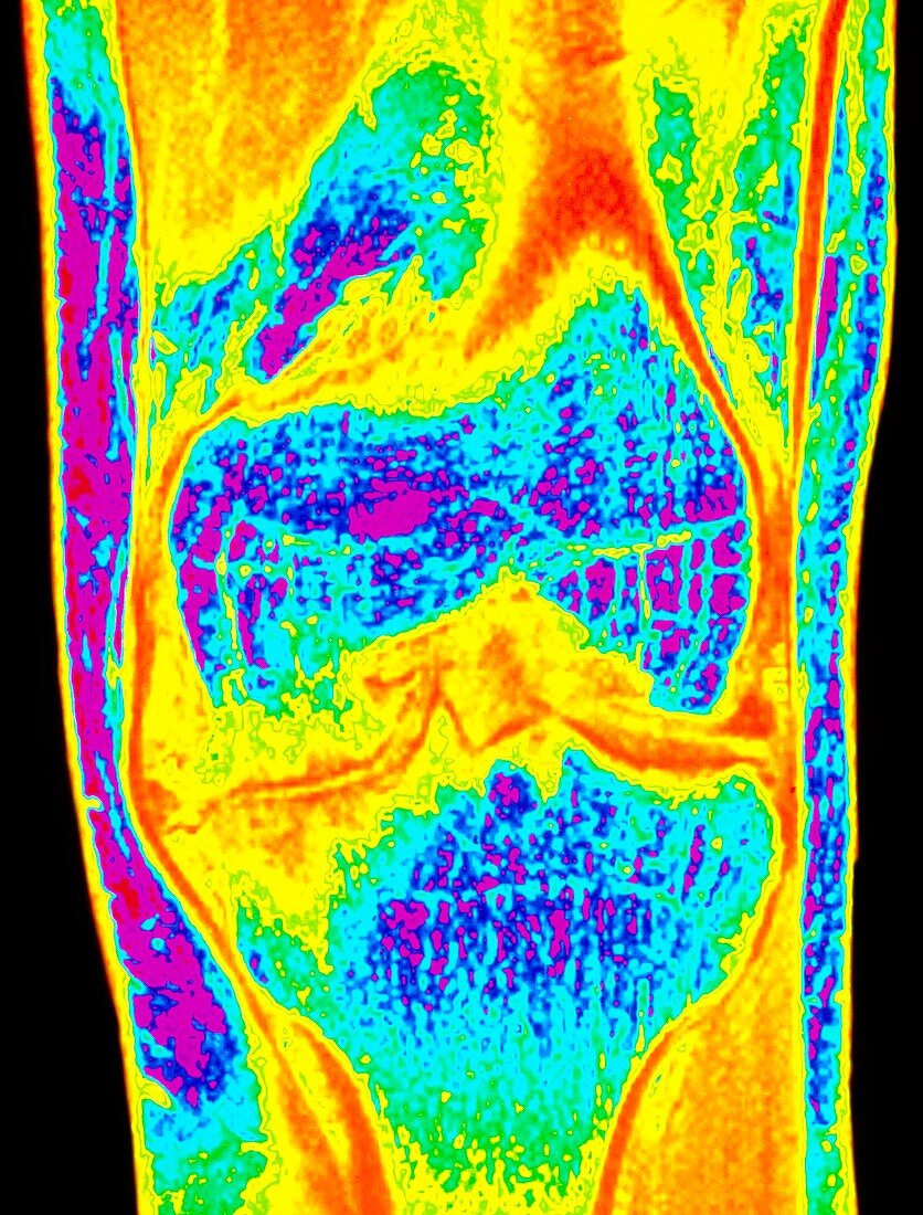
| Osteoarthritis of knee. Colour Magnetic Resonance Imaging (MRI) scan of a front view of the knee joint of a 33 year old male with osteoarthritis. The lower end of the femur (thigh bone) is at centre (blue); it articulates with the upper end of the tibia (shin bone) at lower centre (blue). Due to osteoarthritis the ends of the bones have become severely eroded and jagged; cracking of the bone is also visible. A deep intercondylar notch (triangular) has formed in the femur. Osteo- arthritis is the most common type of arthritis due mainly to wear and tear on joints in the elderly. This patient had haemophilia which was a factor | |
| Licence : | Droits gérés |
| Crédit: | Science Photo Library / SIMON FRASER, ROYAL VICTORIA INFIRMARY |
| Taille de l’image : | 3681 px × 4843 px |
| Model Release : | Non requis |
| Property Release : | Non requis |
| Restrictions : | - |
Prix pour cette image À partir de 45 €
Produit vendu
(Calendrier, Carte postale, Carte de vœux, Impression sur textile, Packaging etc)
À partir de 45 €
Usage commercial
(Affichage, Annonce presse, Annonce TV, Carte, Digital - hors rés. sociaux, Digital - rés. sociaux etc)
À partir de 45 €
Éditorial
(Digital, Journal, Livre, Livre pratique, Magazine, Télévision etc)
À partir de 60 €
Usage non-commercial
(Digital - hors rés. sociaux, Digital - rés. sociaux etc)
À partir de 120 €
Mots clés
- affection articulaire,
- affection des articulations,
- arthrite,
- arthritique,
- arthritis,
- arthrose,
- articulation,
- articulation du genou,
- désordre,
- état,
- genou,
- hémophilie,
- maladie,
- maladie articulaire,
- maladie des articulations,
- médecine,
- médical,
- médicale,
- ostéo-,
- osteoarthritis,
- soins de santé,
- trouble