MRI scan of rheumatoid arthritis of neck
Numéro d’image : 11835029
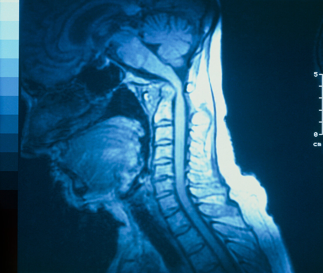
| Rheumatoid arthritis. Magnetic resonance image (MRI) of the neck of a 70 years-old woman suffering from rheumatoid arthritis,showing the subluxation (partial dislocation) of the atlanto- axial joint. This joint between the top two cervical vertebrae (the atlas & axis) may become dangerously weakened in rheumatoid arthritis,due to destruction of the ligaments that bind the joint. As a result,the spinal cord has become compressed,and appears pinched & abnormally bent in this image. Symptoms of shooting pains & weakness in the limbs result; severe cases may produce quadriplegia & even sudden death | |
| Licence : | Droits gérés |
| Crédit: | Science Photo Library / NEWCASTLE GENERAL HOSPITAL / NEURORADIOLOGY DEPT / SIMON FRASER |
| Taille de l’image : | 4760 px × 4026 px |
| Model Release : | Non requis |
| Property Release : | Non requis |
| Restrictions : | - |
Prix pour cette image À partir de 45 €
Produit vendu
(Calendrier, Carte postale, Carte de vœux, Impression sur textile, Packaging etc)
À partir de 45 €
Usage commercial
(Affichage, Annonce presse, Annonce TV, Carte, Digital - hors rés. sociaux, Digital - rés. sociaux etc)
À partir de 45 €
Éditorial
(Digital, Journal, Livre, Livre pratique, Magazine, Télévision etc)
À partir de 60 €
Usage non-commercial
(Digital - hors rés. sociaux, Digital - rés. sociaux etc)
À partir de 120 €