Artwork of inflamed bronchial epithelium in asthma
Numéro d’image : 11834772
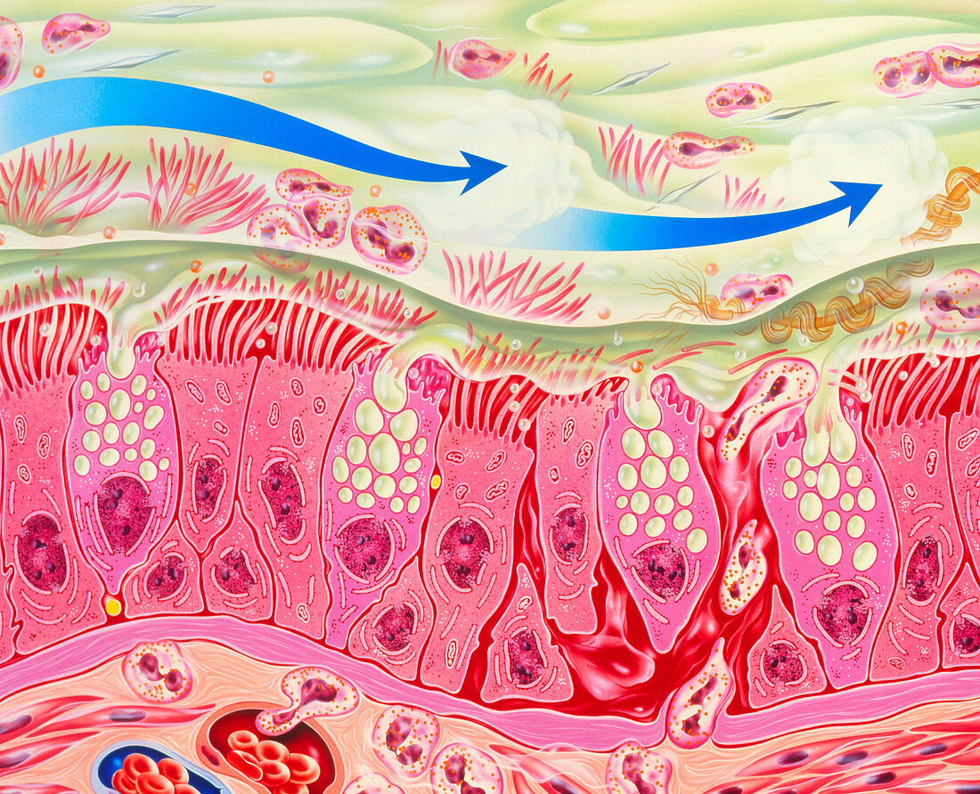
| Bronchial inflammation in asthma. Artwork of bron- chial epithelial tissue inflamed by asthma. A thick layer of mucus (green) covers the tissue. This is produced by goblet cells interspersed among ciliated epithelial cells. The connective tissue at the bottom contains enlarged blood vess- els (lower left) and numerous eosinophil white blood cells,which are also abundant in the mucus. Eosinophils play a major role in allergic inflam- mations. They secrete chemicals that are partly responsible for the bronchial constriction that occurs in asthma. Epithelial desquamation (shed- ding of outer layer of cells) is shown at right. The blue arrows symbolise drugs taken by inhaler | |
| Licence : | Droits gérés |
| Crédit: | Science Photo Library / Bavosi, John |
| Taille de l’image : | 3424 px × 2778 px |
| Model Release : | Non requis |
| Property Release : | Non requis |
| Restrictions : | - |
Prix pour cette image À partir de 45 €
Produit vendu
(Calendrier, Carte postale, Carte de vœux, Impression sur textile, Packaging etc)
À partir de 45 €
Usage commercial
(Affichage, Annonce presse, Annonce TV, Carte, Digital - hors rés. sociaux, Digital - rés. sociaux etc)
À partir de 45 €
Éditorial
(Digital, Journal, Livre, Livre pratique, Magazine, Télévision etc)
À partir de 60 €
Usage non-commercial
(Digital - hors rés. sociaux, Digital - rés. sociaux etc)
À partir de 120 €