Colour TEM of the influenza virus infecting a cell
Numéro d’image : 11833844
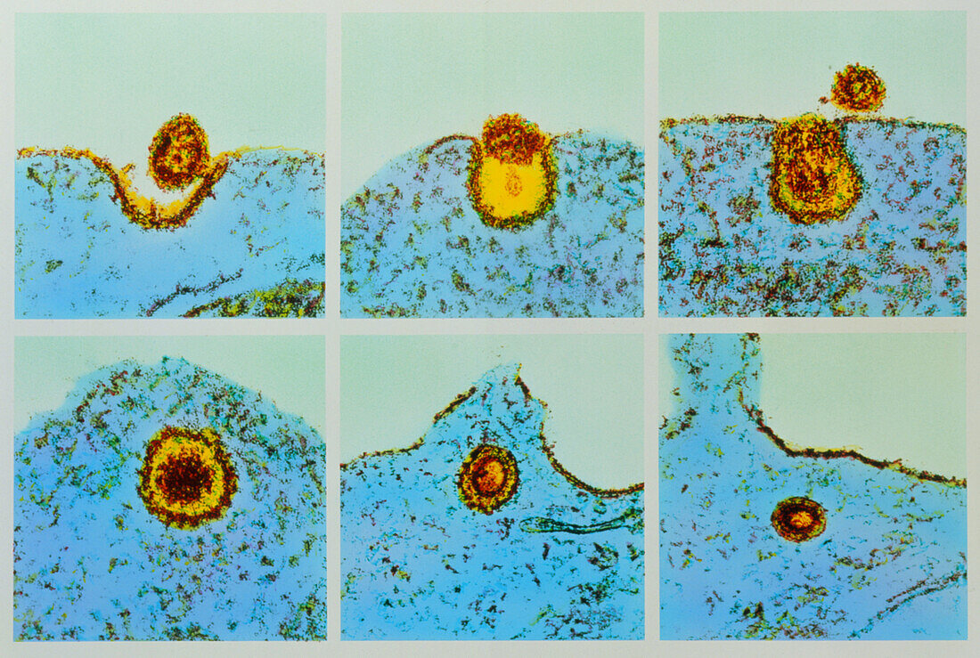
| Flu virus infecting a cell. Coloured Transmission Electron Micrograph of stages of cell infection of an influenza virus. The virus appears rounded in shape,with a core of ribonucleic acid (RNA). It has a spiked outer coat which allows the virus to attach to host cells. Host cell cytoplasm appears granular. At top frames (3 images) the virus attaches to the cell,causing the cell membrane to fold around the virus. At lower frames (3 images) the virus penetrates the cell,infecting it,and causing more influenza viruses to be produced. This virus is contagious and invades mucus cells in the respiratory tract. Magnification: x50,000 (for each inset) at 5x7cm size. x180,000 for each 3x3 inch size inset at 10x8ins | |
| Licence : | Droits gérés |
| Crédit: | Science Photo Library / Patterson, Dr. Steve |
| Taille de l’image : | 3689 px × 2480 px |
| Model Release : | Non requis |
| Property Release : | Non requis |
| Restrictions : | - |
Prix pour cette image À partir de 45 €
Produit vendu
(Calendrier, Carte postale, Carte de vœux, Impression sur textile, Packaging etc)
À partir de 45 €
Usage commercial
(Affichage, Annonce presse, Annonce TV, Carte, Digital - hors rés. sociaux, Digital - rés. sociaux etc)
À partir de 45 €
Éditorial
(Digital, Journal, Livre, Livre pratique, Magazine, Télévision etc)
À partir de 60 €
Usage non-commercial
(Digital - hors rés. sociaux, Digital - rés. sociaux etc)
À partir de 120 €