Cytomegalovirus infection
Numéro d’image : 11833343
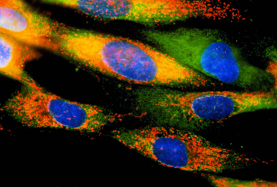
| Cytomegalovirus infected cells. Immunofluorescent light micrograph of human fibroblast culture cells infected with cytomegaloviruses. The infected cells are shown by the presence of the virus- specific protein UL37 (orange/yellow). Cell nuclei are blue,with endoplasmic reticulum green. UL37 localizes to the mitochondria,keeping the cells alive for longer and enabling them to produce more viruses. Fibroblasts are found in human connective tissue. They are involved in,among other things,making the fibrous structural protein collagen. In immunofluorescence,fluorescent dyes are attached to specific tissues using antibodies. Magnification: x200 at 5x7cm size | |
| Licence : | Droits gérés |
| Crédit: | Science Photo Library / Kedersha, Nancy |
| Taille de l’image : | 3673 px × 2480 px |
| Model Release : | Non requis |
| Property Release : | Non requis |
| Restrictions : | - |
Prix pour cette image À partir de 45 €
Produit vendu
(Calendrier, Carte postale, Carte de vœux, Impression sur textile, Packaging etc)
À partir de 45 €
Usage commercial
(Affichage, Annonce presse, Annonce TV, Carte, Digital - hors rés. sociaux, Digital - rés. sociaux etc)
À partir de 45 €
Éditorial
(Digital, Journal, Livre, Livre pratique, Magazine, Télévision etc)
À partir de 60 €
Usage non-commercial
(Digital - hors rés. sociaux, Digital - rés. sociaux etc)
À partir de 120 €