False-col TEM of myxomatosis virus
Numéro d’image : 11832995
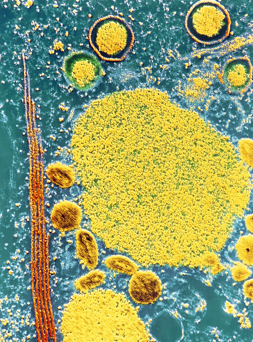
| False-colour transmission electron micrograph (TEM) of a cell affected with the myxomatosis virus (a member of the poxvirus group),showing sites of viral synthesis & replication within the cell & viruses in different stages of evolution. The central yellow area can be considered as the site of viral synthesis & is known as the viroplasma. Viruses are seen in 3 stages of development. The larger circular particles at top (with separate surrounding membrane) are in 1st stage,the smaller,dark oval bodies at bottom left are in 2nd stage,& the nearby dark circular viruses are in the 3rd stage of development. Magnification: X 14,700 at 35mm size | |
| Licence : | Droits gérés |
| Crédit: | Science Photo Library / CNRI |
| Taille de l’image : | 4004 px × 5407 px |
| Model Release : | Non requis |
| Property Release : | Non requis |
| Restrictions : | - |
Prix pour cette image À partir de 45 €
Produit vendu
(Calendrier, Carte postale, Carte de vœux, Impression sur textile, Packaging etc)
À partir de 45 €
Usage commercial
(Affichage, Annonce presse, Annonce TV, Carte, Digital - hors rés. sociaux, Digital - rés. sociaux etc)
À partir de 45 €
Éditorial
(Digital, Journal, Livre, Livre pratique, Magazine, Télévision etc)
À partir de 60 €
Usage non-commercial
(Digital - hors rés. sociaux, Digital - rés. sociaux etc)
À partir de 120 €