An X-ray diffractometer
Numéro d’image : 11826723
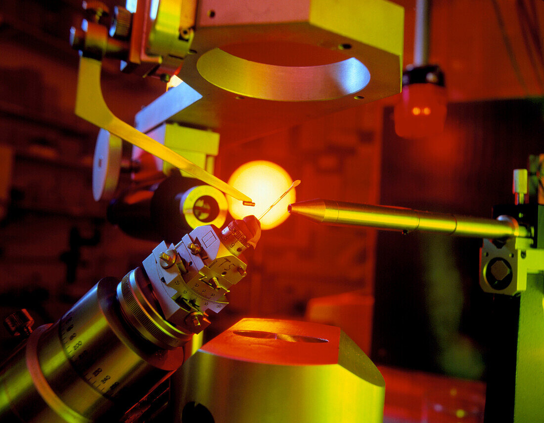
| X-ray diffraction crystallography. A crystal of methanol dehydrogenase is bombarded with X-rays to determine its molecular structure. The dimensions of the crystal sample,visible here in the phial at centre,are usually only around 0.25 x 0.5 millimetres. The repeated pattern of the crystal lattice acts as a diffraction grating,diffracting the beam in a way which depends on the spacing and arrangement of atoms in the lattice. The scattered rays then strike a detector plate; the data is recorded either on X-ray sensitive photographic film,or with electronic equipment linked to a computer | |
| Licence : | Droits gérés |
| Crédit: | Science Photo Library / King-Holmes, James / Ocms |
| Taille de l’image : | 3156 px × 2460 px |
| Model Release : | Non requis |
| Property Release : | Non requis |
| Restrictions : | - |
Prix pour cette image À partir de 45 €
Produit vendu
(Calendrier, Carte postale, Carte de vœux, Impression sur textile, Packaging etc)
À partir de 45 €
Usage commercial
(Affichage, Annonce presse, Annonce TV, Carte, Digital - hors rés. sociaux, Digital - rés. sociaux etc)
À partir de 45 €
Éditorial
(Digital, Journal, Livre, Livre pratique, Magazine, Télévision etc)
À partir de 60 €
Usage non-commercial
(Digital - hors rés. sociaux, Digital - rés. sociaux etc)
À partir de 120 €