An X-ray diffractometer
Numéro d’image : 11826722
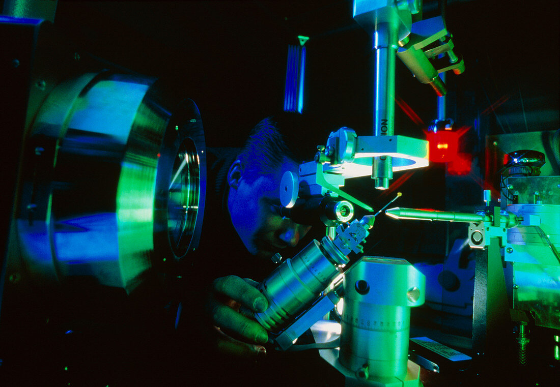
| X-ray diffraction crystallography. A crystal of phosphorylase B is bombarded with X-rays to determine its molecular structure. The crystal sample,contained in the phial at centre,is approximately 1.5 x 0.7 millimetres. The repeated pattern of the crystal lattice acts as a diffrac- tion grating,diffracting the beam in a way which depends on the lattice's arrangement and spacing. The scattered rays then strike a detector. The tube at the top of the picture is a cryogenic device which cools the specimen to 100 kelvin (-173 celsius),ensuring the stability of the crystal for the duration of the experiment | |
| Licence : | Droits gérés |
| Crédit: | Science Photo Library / King-Holmes, James / Ocms |
| Taille de l’image : | 5037 px × 3484 px |
| Model Release : | Non requis |
| Property Release : | Non requis |
| Restrictions : | - |
Prix pour cette image À partir de 45 €
Produit vendu
(Calendrier, Carte postale, Carte de vœux, Impression sur textile, Packaging etc)
À partir de 45 €
Usage commercial
(Affichage, Annonce presse, Annonce TV, Carte, Digital - hors rés. sociaux, Digital - rés. sociaux etc)
À partir de 45 €
Éditorial
(Digital, Journal, Livre, Livre pratique, Magazine, Télévision etc)
À partir de 60 €
Usage non-commercial
(Digital - hors rés. sociaux, Digital - rés. sociaux etc)
À partir de 120 €