Researcher using an X-ray diffractometer
Numéro d’image : 11826720
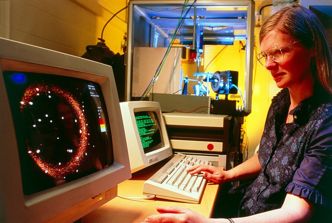
| MODEL RELEASED Researcher using X-ray diffraction crystallography equipment to determine the crystal structure of a protein. A beam of monochromatic X- rays is generated & directed at the crystal (held in the apparatus at back). The repeated pattern of the crystal lattice acts as a diffraction grating,diffracting the beam in a way which depends on the lattice's arrangement and spacing. The scattered rays then strike a detector plate; the intensity at each point is recorded on X-ray sensitive photographic film,or else,as here,by electronic equipment which digitises the data for analysis & presentation on a computer (see monitor on left) | |
| Licence : | Droits gérés |
| Crédit: | Science Photo Library / King-Holmes, James / Ocms |
| Taille de l’image : | 5101 px × 3424 px |
| Model Release : | Disponible |
| Property Release : | Non requis |
| Restrictions : | - |
Prix pour cette image À partir de 45 €
Produit vendu
(Calendrier, Carte postale, Carte de vœux, Impression sur textile, Packaging etc)
À partir de 45 €
Usage commercial
(Affichage, Annonce presse, Annonce TV, Carte, Digital - hors rés. sociaux, Digital - rés. sociaux etc)
À partir de 45 €
Éditorial
(Digital, Journal, Livre, Livre pratique, Magazine, Télévision etc)
À partir de 60 €
Usage non-commercial
(Digital - hors rés. sociaux, Digital - rés. sociaux etc)
À partir de 120 €