LM of cytoskeletons
Numéro d’image : 11821560
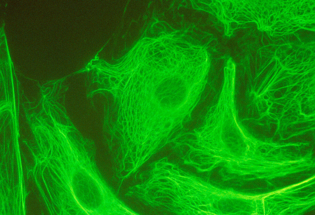
| Ultraviolet fluorescence light micrograph of cytoskeletons showing the distribution of an intermediate filament protein called vimentin. The cells are from the kidney epithelium of the kangaroo-rat. The cytoskeleton falls into two components; microtubules,which are rigid protein tubes that act as direction markers in a cell and two filamentous components called microfilaments & intermediate filaments. Intermediate filaments,are flexible,forming three dimensional networks. They are thought to be directly involved in the movement of organelles,vesicles & membranes. The oval depressions seen here are the sites of the nuclei. Magnification x100 at 35mm size. Microcosmos P 118 fig.6.17 | |
| Licence : | Droits gérés |
| Crédit: | Science Photo Library / Dawson, Dr. Peter |
| Taille de l’image : | 5140 px × 3510 px |
| Model Release : | Non requis |
| Property Release : | Non requis |
| Restrictions : | - |
Prix pour cette image À partir de 45 €
Produit vendu
(Calendrier, Carte postale, Carte de vœux, Impression sur textile, Packaging etc)
À partir de 45 €
Usage commercial
(Affichage, Annonce presse, Annonce TV, Carte, Digital - hors rés. sociaux, Digital - rés. sociaux etc)
À partir de 45 €
Éditorial
(Digital, Journal, Livre, Livre pratique, Magazine, Télévision etc)
À partir de 60 €
Usage non-commercial
(Digital - hors rés. sociaux, Digital - rés. sociaux etc)
À partir de 120 €