Cell microtubules/cytoskeleton
Numéro d’image : 11821557
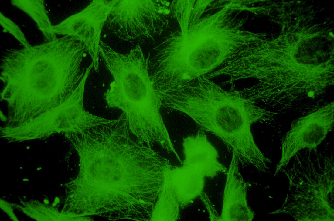
| False-colour tubulin immunofluorescence micrograph of tissue culture cells showing the microtubule component of the cytoskeleton,the three- dimensional array of protein molecules that permeates the cytoplasm of a cell. Here,the cells have been fixed and stained with an antibody which binds to the microtubules; the antibody is labelled with the dye fluorescein,which fluoresces green in ultraviolet light. The apparently empty space in the centre of each cell represents the nucleus. Microtubules are thought to act as direction-markers within the cell - a sort of cytoplasmic railway network. Magnification: x275 at 35mm size. Dark green tint | |
| Licence : | Droits gérés |
| Crédit: | Science Photo Library / Murti, Dr. Gopal |
| Taille de l’image : | 3737 px × 2480 px |
| Model Release : | Non requis |
| Property Release : | Non requis |
| Restrictions : | - |
Prix pour cette image À partir de 45 €
Produit vendu
(Calendrier, Carte postale, Carte de vœux, Impression sur textile, Packaging etc)
À partir de 45 €
Usage commercial
(Affichage, Annonce presse, Annonce TV, Carte, Digital - hors rés. sociaux, Digital - rés. sociaux etc)
À partir de 45 €
Éditorial
(Digital, Journal, Livre, Livre pratique, Magazine, Télévision etc)
À partir de 60 €
Usage non-commercial
(Digital - hors rés. sociaux, Digital - rés. sociaux etc)
À partir de 120 €