Colour SEM of endoplasmic reticulum in Leydig cell
Numéro d’image : 11821437
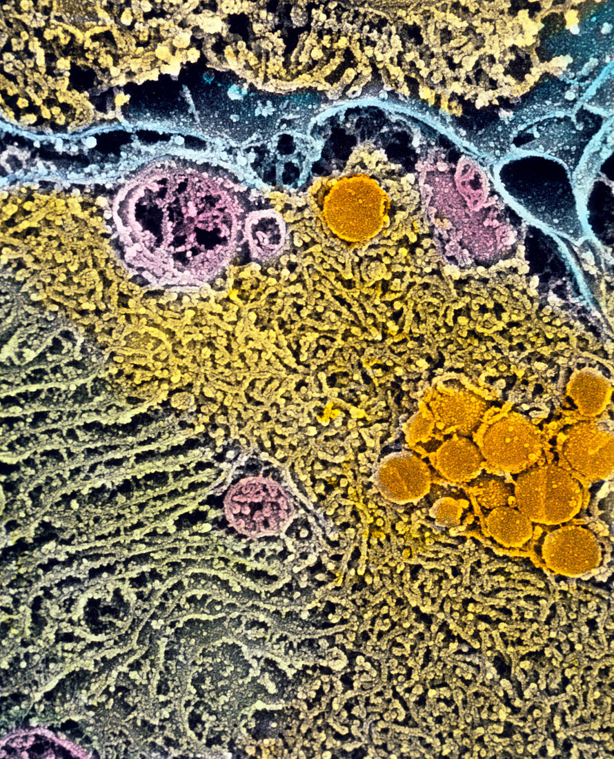
| Leydig cell of testis. Coloured Scanning Electron Micrograph (SEM) of endoplasmic reticulum in a Leydig cell of a 14 week human foetus. Leydig cells produce steroids in the male testis. Here,endoplasmic reticulum (ER) membranes fill most of this secretory cell. Smooth ER is yellow; rough ER (green) is dotted with ribosomes involved in protein synthesis. The synthesis of lipids and steroids occurs within the lumen of these paired ER membranes. Lipid droplets are the round orange structures. Mitochondria (pink) produce chemical energy; the cell membrane is coloured blue. Magnification: x8,100 at 6x7cm size. x10,400 at 4x5ins | |
| Licence : | Droits gérés |
| Crédit: | Science Photo Library / PROFESSORS P.M. MOTTA, S. MAKABE & T. NAGURO |
| Taille de l’image : | 2865 px × 3543 px |
| Model Release : | Non requis |
| Property Release : | Non requis |
| Restrictions : | - |
Prix pour cette image À partir de 45 €
Produit vendu
(Calendrier, Carte postale, Carte de vœux, Impression sur textile, Packaging etc)
À partir de 45 €
Usage commercial
(Affichage, Annonce presse, Annonce TV, Carte, Digital - hors rés. sociaux, Digital - rés. sociaux etc)
À partir de 45 €
Éditorial
(Digital, Journal, Livre, Livre pratique, Magazine, Télévision etc)
À partir de 60 €
Usage non-commercial
(Digital - hors rés. sociaux, Digital - rés. sociaux etc)
À partir de 120 €