Freeze fracture micrograph of cell nucleus
Numéro d’image : 11821391
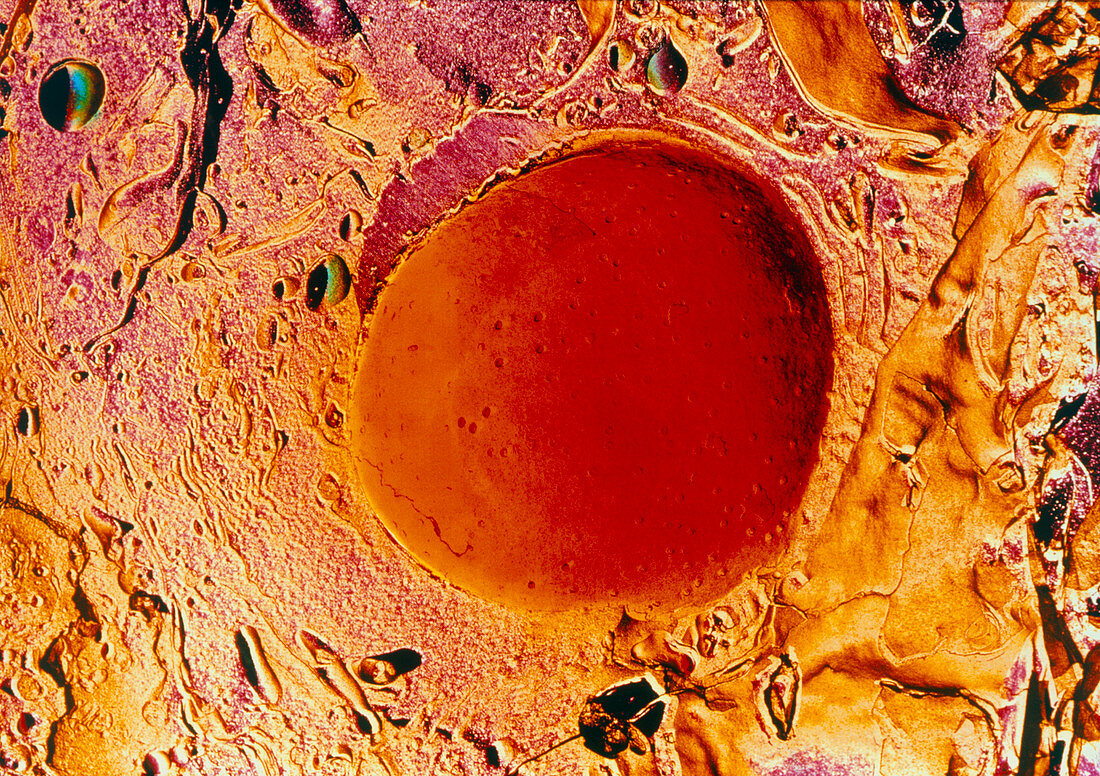
| Cell nucleus. Freeze-fracture scanning electron micrograph of the nucleus (red) and surrounding cytoplasm in parathyroid gland cell. The small pits on the nucleus are pores in its double membrane. These allow large molecules to pass between the nucleus and the cell cytoplasm. This image was made by freezing the cell,fracturing it in a vacuum and then coating the sample with metal or carbon. The metal or carbon replica was then photographed with an electron microscope. The parathyroid glands are pea-shaped glands in the neck that control the level of calcium in the blood. Magnification: x5200 at 7x5cm size. Magnification x2600 at 35mm size | |
| Licence : | Droits gérés |
| Crédit: | Science Photo Library / PROF S. CINTI |
| Taille de l’image : | 4913 px × 3468 px |
| Model Release : | Non requis |
| Property Release : | Non requis |
| Restrictions : | - |
Prix pour cette image À partir de 45 €
Produit vendu
(Calendrier, Carte postale, Carte de vœux, Impression sur textile, Packaging etc)
À partir de 45 €
Usage commercial
(Affichage, Annonce presse, Annonce TV, Carte, Digital - hors rés. sociaux, Digital - rés. sociaux etc)
À partir de 45 €
Éditorial
(Digital, Journal, Livre, Livre pratique, Magazine, Télévision etc)
À partir de 60 €
Usage non-commercial
(Digital - hors rés. sociaux, Digital - rés. sociaux etc)
À partir de 120 €