Immunfluorescent LM of cell death (apoptosis)
Numéro d’image : 11821336
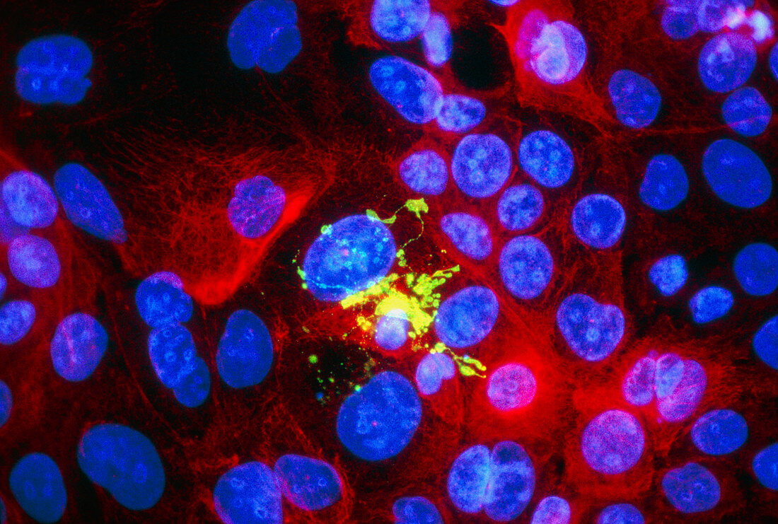
| Apoptosis. Immunofluorescent Light Micrograph of a culture of normal human epithelial cells,showing apoptosis or "genetically programmed cell death". Cell nuclei are blue; cytoplasm stains red or green. At centre a single green cell has undergone apoptosis: its nucleus & cytoplasm have fragmented and shrunk into blobs which sometimes resemble grape-like clusters. These cell remnants will then be eaten by the neighbouring cells. Research on apoptosis may provide genetic treatments for diseases such as cancer. Immunofluorescence uses antibodies to attach fluorescent dyes to cell structures. Magnification: x250 at 5x7cm size. x125 at 35mm | |
| Licence : | Droits gérés |
| Crédit: | Science Photo Library / Kedersha, Nancy |
| Taille de l’image : | 5226 px × 3522 px |
| Model Release : | Non requis |
| Property Release : | Non requis |
| Restrictions : | - |
Prix pour cette image À partir de 45 €
Produit vendu
(Calendrier, Carte postale, Carte de vœux, Impression sur textile, Packaging etc)
À partir de 45 €
Usage commercial
(Affichage, Annonce presse, Annonce TV, Carte, Digital - hors rés. sociaux, Digital - rés. sociaux etc)
À partir de 45 €
Éditorial
(Digital, Journal, Livre, Livre pratique, Magazine, Télévision etc)
À partir de 60 €
Usage non-commercial
(Digital - hors rés. sociaux, Digital - rés. sociaux etc)
À partir de 120 €
Mots clés
- apoptose,
- apoptosis,
- biologie,
- biologique,
- cellule,
- cytologie,
- décès,
- fluorescent,
- immunofluorescence,
- microbiologie,
- micrographe optique confocal,
- microscope optique,
- microscope optique confocal,
- microscopie de fluorescence,
- microscopie optique,
- microscopie par fluorescence,
- mort cellulaire,
- structure cellulaire