Cultured cell,light micrograph
Numéro d’image : 11821043
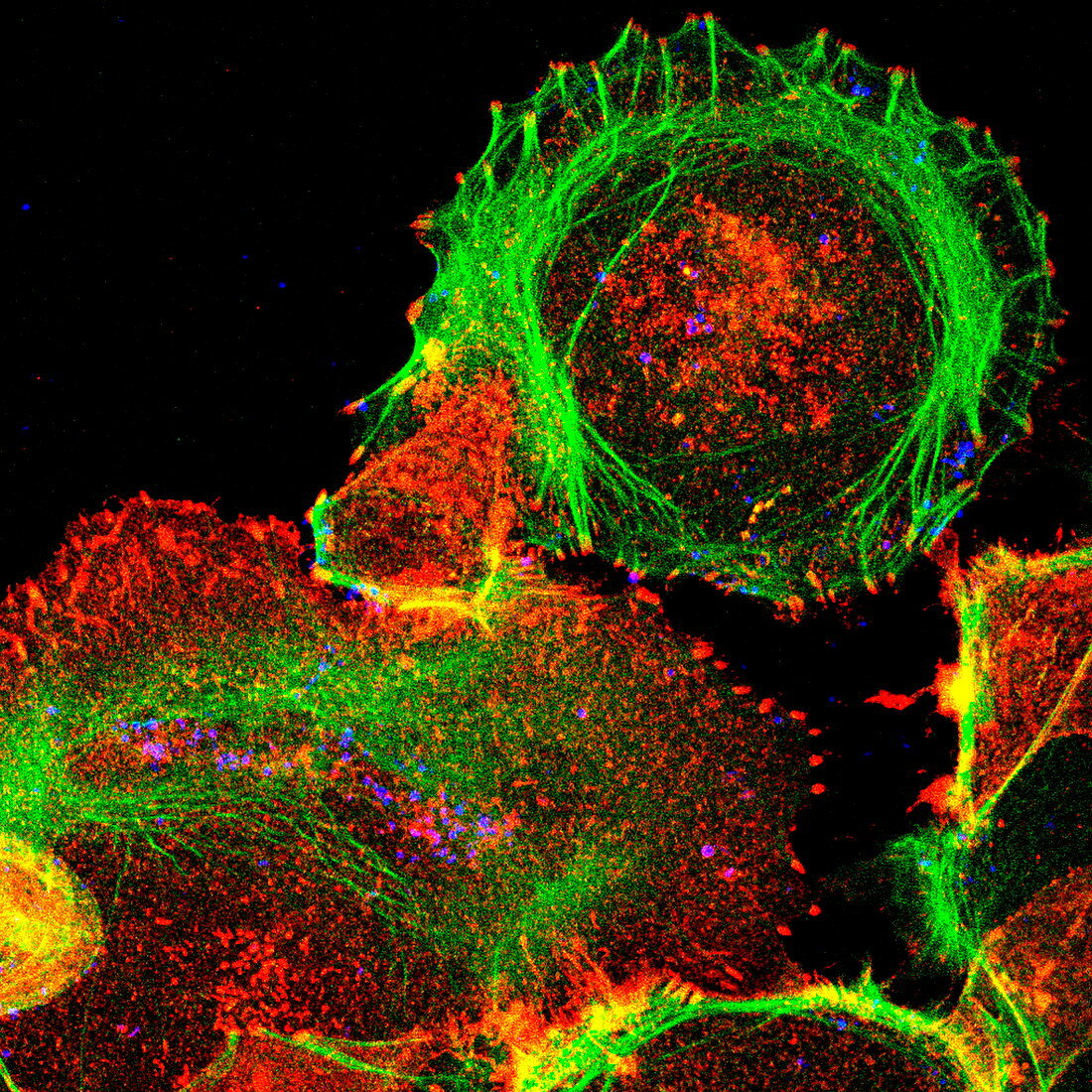
| Cultured cell. Immunofluorescent light micrograph of an epithelial cell (round,upper right). The cell proteins are marked by fluorescent dyes. The actin cytoskeleton is green,bacteria are the blue dots,and red marks an activated protein called phosphotyrosine (a protein that plays a role in the regulation of cell growth). The actin fibres surrounding the cell at upper right end in areas of this activated protein. Yellow marks an overlap between green and red. Epithelial cells make up the body's internal and external surface layers,such as the skin and the walls of the intestines | |
| Licence : | Droits gérés |
| Crédit: | Science Photo Library |
| Taille de l’image : | 2929 px × 2929 px |
| Model Release : | Non requis |
| Property Release : | Non requis |
| Restrictions : | - |
Prix pour cette image À partir de 45 €
Produit vendu
(Calendrier, Carte postale, Carte de vœux, Impression sur textile, Packaging etc)
À partir de 45 €
Usage commercial
(Affichage, Annonce presse, Annonce TV, Carte, Digital - hors rés. sociaux, Digital - rés. sociaux etc)
À partir de 45 €
Éditorial
(Digital, Journal, Livre, Livre pratique, Magazine, Télévision etc)
À partir de 60 €
Usage non-commercial
(Digital - hors rés. sociaux, Digital - rés. sociaux etc)
À partir de 120 €
Mots clés
- actine,
- bacteria,
- bactérie,
- bacterium,
- biologie,
- biologique,
- cellulaire,
- cellule,
- croissance cellulaire,
- cytologie,
- cytologique,
- en bonne santé,
- épithéliale,
- epithelium,
- épithélium,
- fluorescent,
- histologie,
- histologique,
- immunofluorescence,
- micrographie optique,
- microscope optique,
- microscopie optique,
- normal,
- sain