Kidney cells
Numéro d’image : 11821036
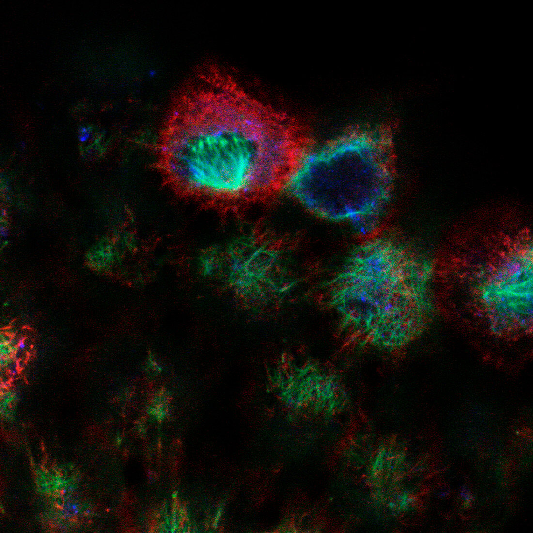
| Kidney cells. Confocal fluorescence light micrograph of cultured kidney cells. Antibody- linked dyes have been used to stain tubulin (green),actin (red) and Golgi protein (blue). Tubulin and actin are proteins of the cytoskeleton,the internal framework of a cell involved in cell movement and transport of substances. Golgi proteins are part of the Golgi apparatus,a system of compartments involved in sorting cell products. A confocal microscope only detects light from the focal point of its objective lens. By moving the focal point,images of thin sections of an intact specimen can be obtained. Magnification unknown | |
| Licence : | Droits gérés |
| Crédit: | Science Photo Library / Reichelt, Stefanie |
| Taille de l’image : | 1024 px × 1024 px |
| Model Release : | Non requis |
| Property Release : | Non requis |
| Restrictions : | - |
Prix pour cette image À partir de 45 €
Produit vendu
(Calendrier, Carte postale, Carte de vœux, Impression sur textile, Packaging etc)
À partir de 45 €
Usage commercial
(Affichage, Annonce presse, Annonce TV, Carte, Digital - hors rés. sociaux, Digital - rés. sociaux etc)
À partir de 45 €
Éditorial
(Digital, Journal, Livre, Livre pratique, Magazine, Télévision etc)
À partir de 60 €
Usage non-commercial
(Digital - hors rés. sociaux, Digital - rés. sociaux etc)
À partir de 120 €
Mots clés
- actine,
- apparatus,
- appareil,
- biologie,
- biologique,
- cellule,
- cellules,
- confocal,
- cultivé,
- culture,
- cytosquelette,
- fluorescence,
- fluorescent,
- Golgi,
- immunofluorescence,
- interphase,
- microbiologie,
- micrographie optique,
- microscope,
- microscope optique,
- microscopie,
- microscopie optique,
- protéine,
- protéines,
- rein,
- tubuline