Coloured LM of HeLa cancer cells
Numéro d’image : 11820989
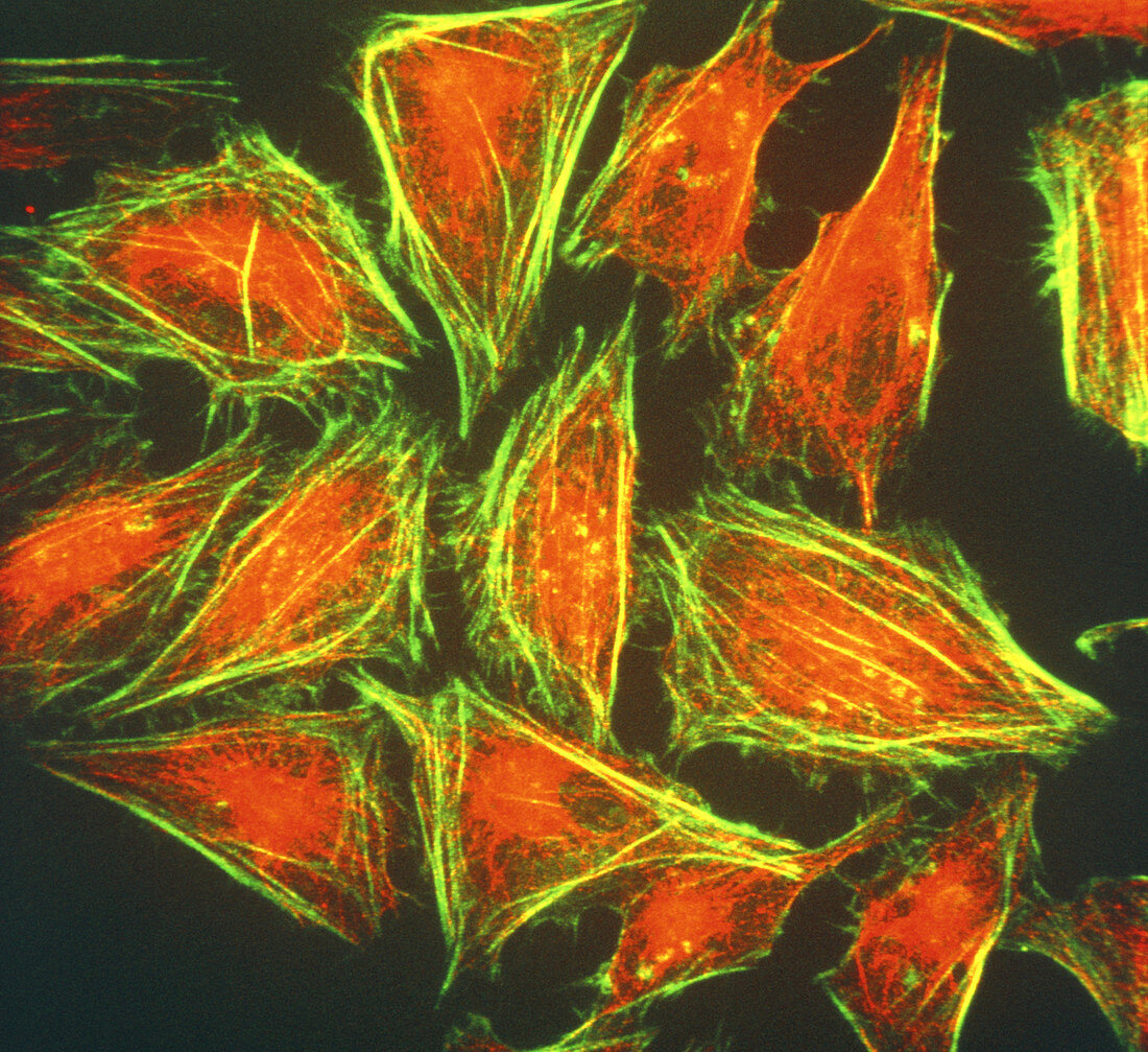
| HeLa cells. Coloured immunofluorescent light micrograph (LM) of HeLa cancer cells. The triangular cells have been stained to reveal the microtubules (red) and actin filaments (green) of the cytoskeleton. HeLa cells were established in 1952 as the first human cell line to research cancer. The cells were named after Henrietta Lacks,from whose cervix they were obtained. HeLa cancer cells are used worldwide as they grow unusually well in the laboratory. Immunofluorescence is a staining technique which uses antibodies to attach fluorescent dyes to specific cell tissues. Magnification: x740 at 6x4.5cm size. x400 at 35mm | |
| Licence : | Droits gérés |
| Crédit: | Science Photo Library / Murti, Dr. Gopal |
| Taille de l’image : | 4779 px × 4377 px |
| Model Release : | Non requis |
| Property Release : | Non requis |
| Restrictions : | - |
Prix pour cette image À partir de 45 €
Produit vendu
(Calendrier, Carte postale, Carte de vœux, Impression sur textile, Packaging etc)
À partir de 45 €
Usage commercial
(Affichage, Annonce presse, Annonce TV, Carte, Digital - hors rés. sociaux, Digital - rés. sociaux etc)
À partir de 45 €
Éditorial
(Digital, Journal, Livre, Livre pratique, Magazine, Télévision etc)
À partir de 60 €
Usage non-commercial
(Digital - hors rés. sociaux, Digital - rés. sociaux etc)
À partir de 120 €
Mots clés
- actine,
- biologie,
- biologique,
- cellule,
- cellule cancéreuse,
- cellule HeLa,
- cellules,
- cellules HeLa,
- cultivé,
- culture,
- culture cellulaire,
- culture de cellule,
- fluorescent,
- hela,
- immunofluorescence,
- microbiologie,
- micrographe optique confocal,
- microscope optique,
- microscope optique confocal,
- microscopie de fluorescence,
- microscopie optique,
- microscopie par fluorescence,
- microtubule