Immunofluorescent LM of HeLa cancer cells
Numéro d’image : 11820987
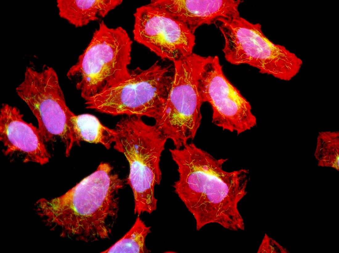
| HeLa cancer cells. Immunofluorescent light micrograph of cultured HeLa cancer cells. The nucleus of each cell is pink,cytoplasm is stained red and green fibres of the protein tubulin are seen around each nucleus. Tubulin is used during cell division to divide chromosomes. HeLa cells are the first human cell line,established in 1952 to research cancer. They were obtained from the cervix of Henrietta Lacks,who died of cervical cancer. HeLa cancer cells are used in research worldwide. Immunofluorescence is a staining technique which uses antibodies to attach fluorescent dyes to specific molecules within cells. Magnification: x125 at 6x4.5cm size | |
| Licence : | Droits gérés |
| Crédit: | Science Photo Library / Kedersha, Nancy |
| Taille de l’image : | 4850 px × 3629 px |
| Model Release : | Non requis |
| Property Release : | Non requis |
| Restrictions : | - |
Prix pour cette image À partir de 45 €
Produit vendu
(Calendrier, Carte postale, Carte de vœux, Impression sur textile, Packaging etc)
À partir de 45 €
Usage commercial
(Affichage, Annonce presse, Annonce TV, Carte, Digital - hors rés. sociaux, Digital - rés. sociaux etc)
À partir de 45 €
Éditorial
(Digital, Journal, Livre, Livre pratique, Magazine, Télévision etc)
À partir de 60 €
Usage non-commercial
(Digital - hors rés. sociaux, Digital - rés. sociaux etc)
À partir de 120 €