3D CT scans of obese laboratory mouse
Numéro d’image : 11820520
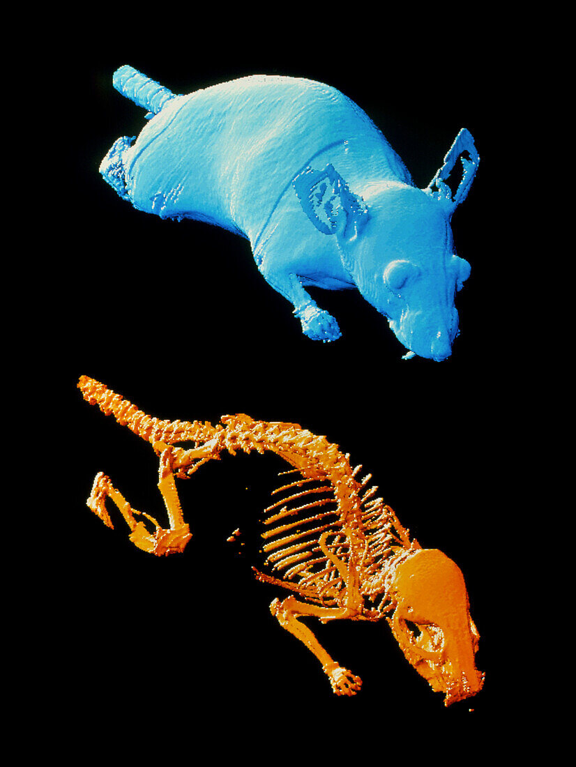
| Mouse CT scans. Coloured three-dimensional (3-D) computed tomography (CT) scans of an obese laboratory mouse (Mus musculus). The skeleton (orange) of this mutant mouse (blue) is at lower frame. This image is from MicroCAT,a CT scanner developed to study small animals like mice by creating images with 10 times the resolution of conventional scanners. The mouse's development can be monitored; mice such as this are bred for genetic analysis. Here the CT scans,slice images through the body made using X-rays,have been built into 3-D images with a computer. The scanner was developed at the Department of Energy's Oak Ridge National Laboratory,USA | |
| Licence : | Droits gérés |
| Crédit: | Science Photo Library / ORNL |
| Taille de l’image : | 2660 px × 3543 px |
| Model Release : | Non requis |
| Property Release : | Non requis |
| Restrictions : | - |
Prix pour cette image À partir de 45 €
Produit vendu
(Calendrier, Carte postale, Carte de vœux, Impression sur textile, Packaging etc)
À partir de 45 €
Usage commercial
(Affichage, Annonce presse, Annonce TV, Carte, Digital - hors rés. sociaux, Digital - rés. sociaux etc)
À partir de 45 €
Éditorial
(Digital, Journal, Livre, Livre pratique, Magazine, Télévision etc)
À partir de 60 €
Usage non-commercial
(Digital - hors rés. sociaux, Digital - rés. sociaux etc)
À partir de 120 €