X-ray diff. pattern formed by an octama of DNA
Numéro d’image : 11817730
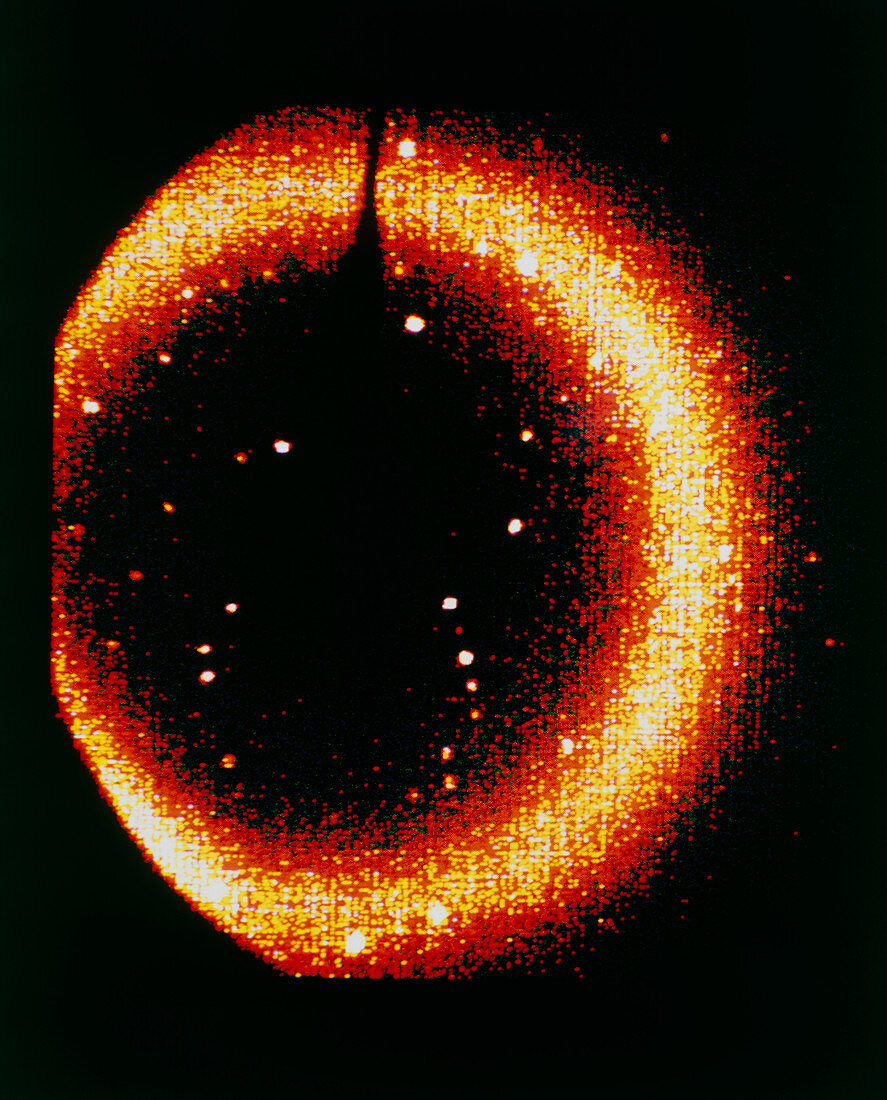
| X-ray diffraction crystallography pattern of DNA. One of a series of screen images showing X-ray diffraction patterns from a crystal formed from a small section of DNA containing 8 base pairs. Complex computer programs are used to produce from the images an electron density map of the crystal,which can then be used to determine the molecular structure. This information is useful for the precise tailoring of drugs for prospective cancer treatments. The white spots are concentrations of diffracted X-rays,and the diffuse circular pattern is background 'noise' from residual water in the crystal | |
| Licence : | Droits gérés |
| Crédit: | Science Photo Library / King-Holmes, James / Ocms |
| Taille de l’image : | 4113 px × 5101 px |
| Model Release : | Non requis |
| Property Release : | Non requis |
| Restrictions : | - |
Prix pour cette image À partir de 45 €
Produit vendu
(Calendrier, Carte postale, Carte de vœux, Impression sur textile, Packaging etc)
À partir de 45 €
Usage commercial
(Affichage, Annonce presse, Annonce TV, Carte, Digital - hors rés. sociaux, Digital - rés. sociaux etc)
À partir de 45 €
Éditorial
(Digital, Journal, Livre, Livre pratique, Magazine, Télévision etc)
À partir de 60 €
Usage non-commercial
(Digital - hors rés. sociaux, Digital - rés. sociaux etc)
À partir de 120 €