F/col STM image of DNA
Numéro d’image : 11817721
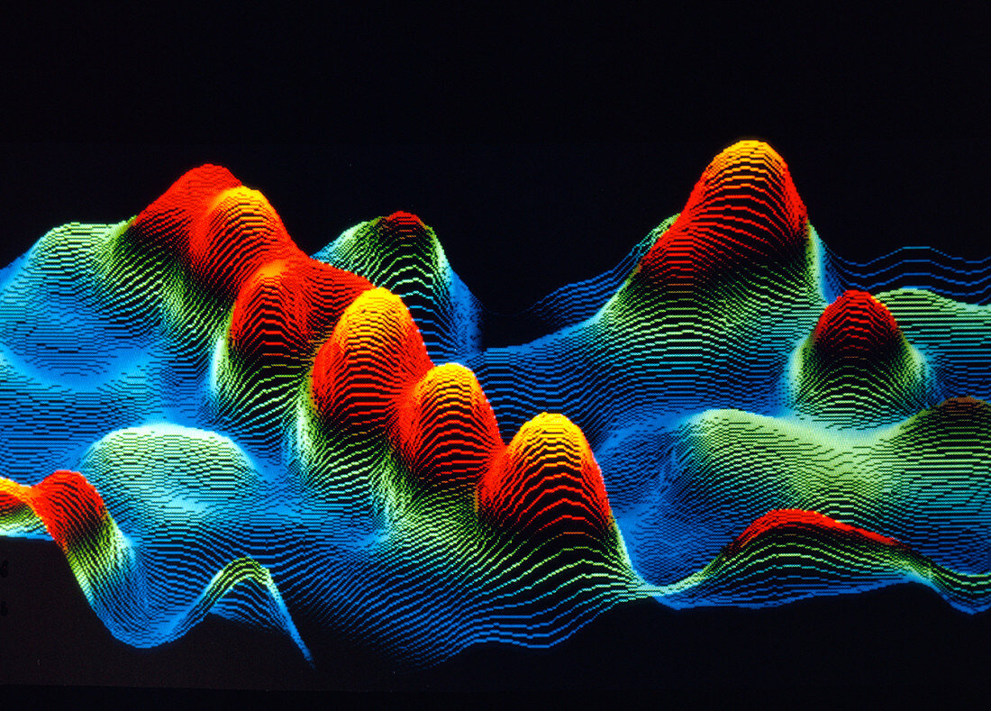
| False-colour scanning tunnelling micrograph (STM) of DNA. A sample of uncoated,double-stranded DNA was dissolved in a salt solution & deposited on graphite prior to being imaged in air by the STM. An STM image is formed by scanning a fine point just above the specimen surface & electronically recording the height of the point as it moves. The main feature of this image is a right-handed,double-stranded DNA molecule (a DNA duplex),which appears as the row of orange/yellow peaks at centre-left. These peaks correspond to the ridges of the DNA double helix. Magnification: x1,600,000 at 6x7cm size | |
| Licence : | Droits gérés |
| Crédit: | Science Photo Library / LAWRENCE LIVERMORE LABORATORY |
| Taille de l’image : | 3543 px × 2540 px |
| Model Release : | Non requis |
| Property Release : | Non requis |
| Restrictions : | - |
Prix pour cette image À partir de 45 €
Produit vendu
(Calendrier, Carte postale, Carte de vœux, Impression sur textile, Packaging etc)
À partir de 45 €
Usage commercial
(Affichage, Annonce presse, Annonce TV, Carte, Digital - hors rés. sociaux, Digital - rés. sociaux etc)
À partir de 45 €
Éditorial
(Digital, Journal, Livre, Livre pratique, Magazine, Télévision etc)
À partir de 60 €
Usage non-commercial
(Digital - hors rés. sociaux, Digital - rés. sociaux etc)
À partir de 120 €