False-colour 3-D CT image of abdominal aorta
Numéro d’image : 11753376
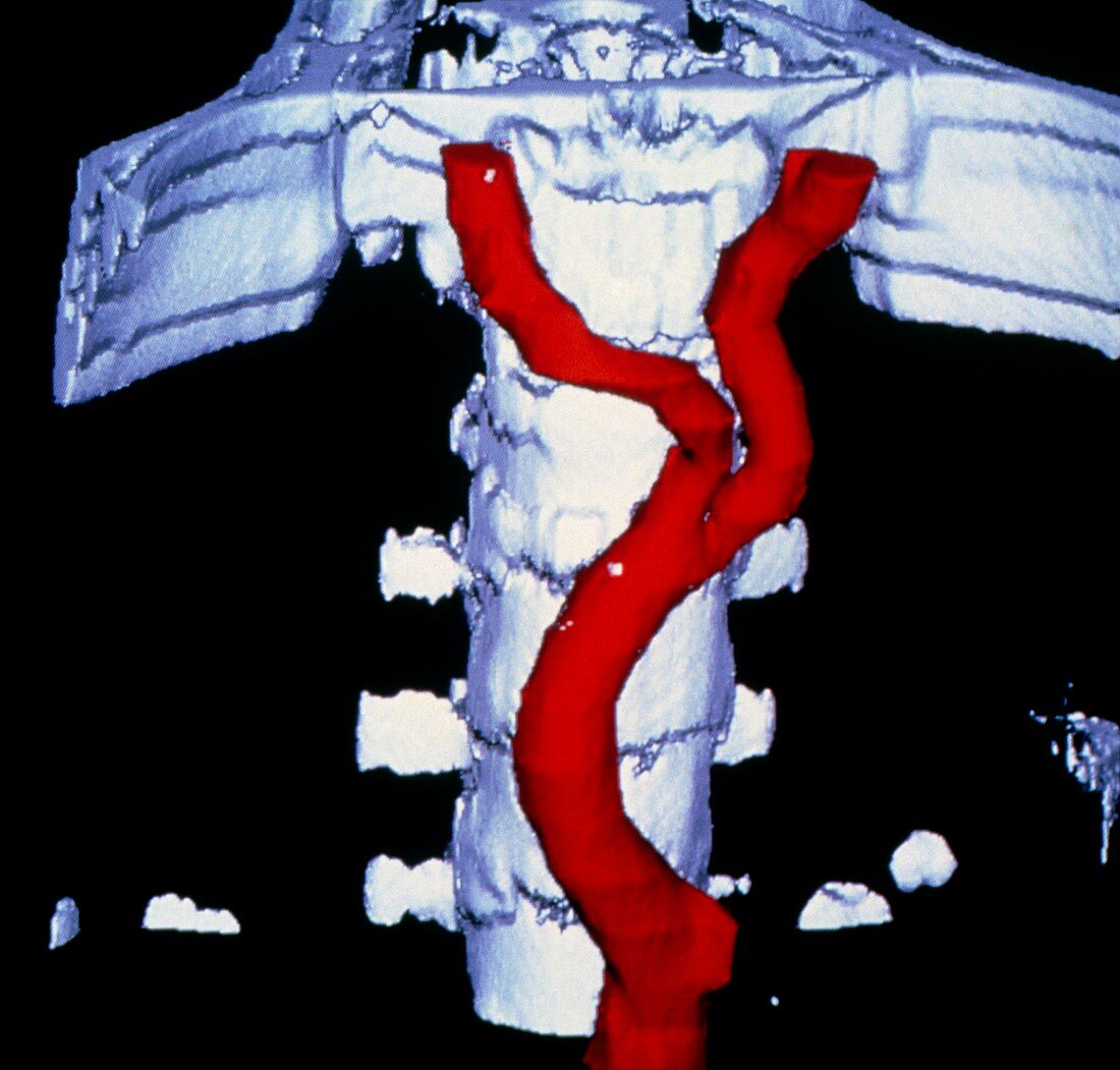
| False-colour three-dimensional image of the division of the abdominal aorta into the iliac arteries,assembled from conventional computed tomography (CT) scan data. Lumbar vertebrae and the upper part of the pelvis appear in white. The aorta & iliac arteries,which serve the legs,are in red. The region of the aorta above the division is liable to various forms of disorders,such as aneurysms & atheroma plaque formation. Imagery of this type might be used to examine the extent of any lesion & in planning surgical repair | |
| Licence : | Libre de droits |
| Crédit: | Science Photo Library / CNRI |
| Model Release : | Non requis |
| Property Release : | Non requis |
| Restrictions : | - |
Prix pour cette image À partir de 29 €
Pour une utilisation numérique (72 dpi)
À partir de 29 €
Pour un usage d'impression (300 dpi)
À partir de 325 €