Blood vessel tumour
Numéro d’image : 11751965
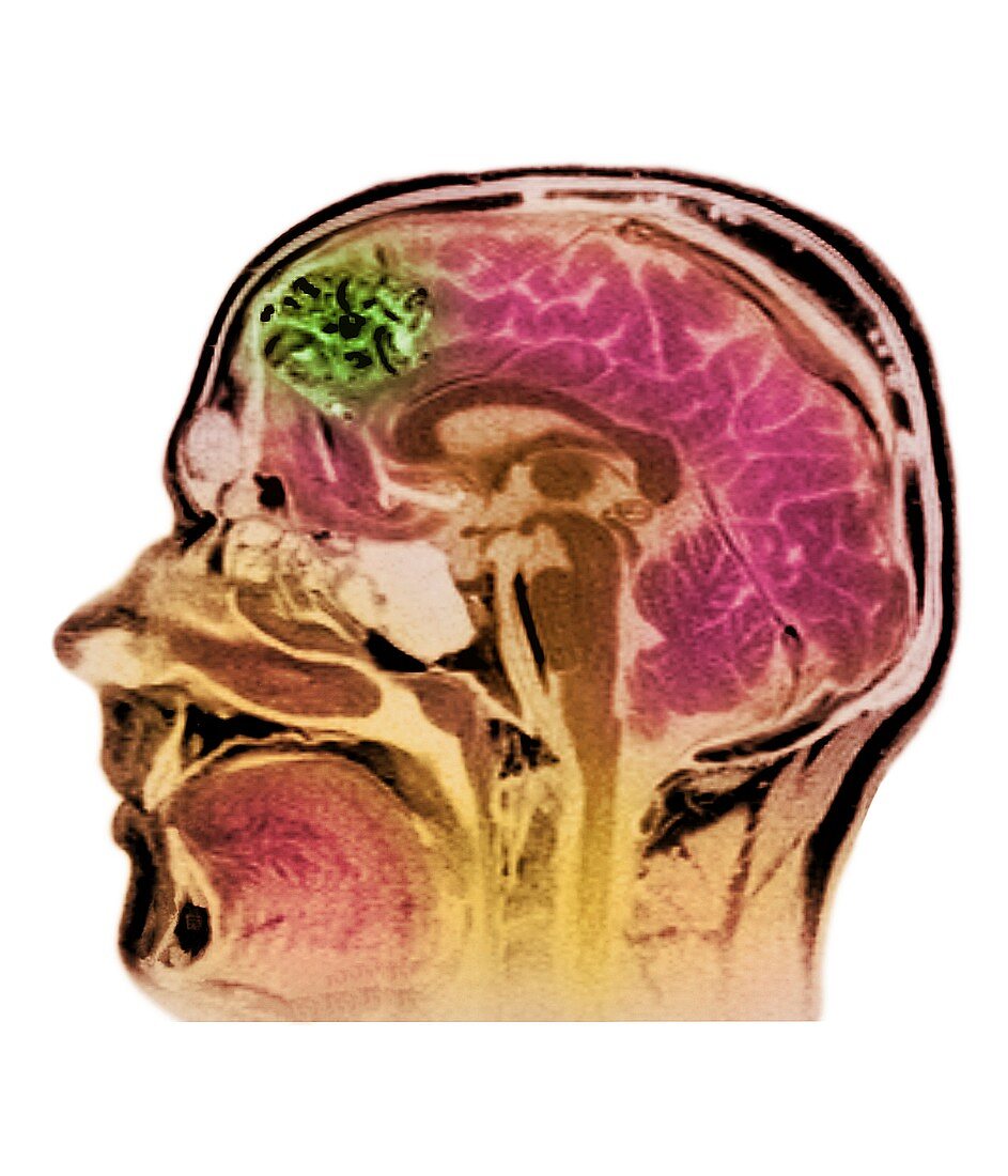
| Blood vessel tumour. Coloured magnetic resonance imaging (MRI) head scan showing an arteriovenous malformation (AVM,green). The AVM is in the brain (mainly red & dark brown) of a 20-year-old patient in this sagittal (vertical) image. This benign (non-cancerous) tumour,also known as an arteriovenous angioma,is formed from a clump of distended blood vessels. It may compress the brain,resulting in epilepsy,or cause a haemorrhage if a vessel bursts. AVMs are surgically removed or destroyed by radiotherapy. MRI scans produce slice images through tissues using a powerful magnet and pulses of radio waves | |
| Licence : | Libre de droits |
| Crédit: | Science Photo Library / RNC, NEWCASTLE / SIMON FRASER |
| Model Release : | Non requis |
| Property Release : | Non requis |
| Restrictions : | - |
Prix pour cette image À partir de 29 €
Pour une utilisation numérique (72 dpi)
À partir de 29 €
Pour un usage d'impression (300 dpi)
À partir de 325 €
Mots clés
- angiome,
- artère,
- artères,
- cerveau,
- désordre,
- diagnostic,
- diagnostique,
- dilaté,
- état,
- hémangiome,
- imagerie par résonance magnétique,
- IRM couleur,
- maladie,
- malformation artérioveineuse,
- médecine,
- médical,
- médicale,
- neurologie,
- neurologique,
- sagittal,
- sagittale,
- soins de santé,
- tête,
- trouble,
- tumeur bénigne,
- vaisseau sanguin,
- vasculaire,
- veines