Staphanoceros rotifer,LM
Numéro d’image : 11734766
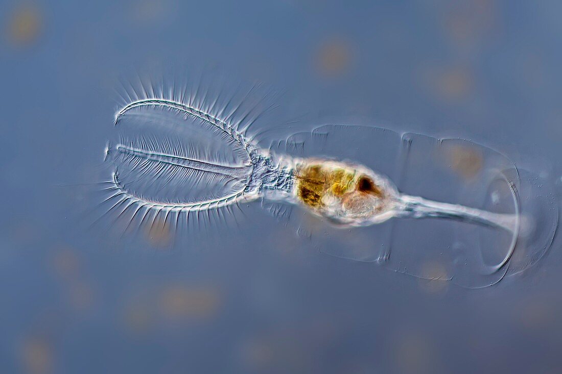
| Light micrograph of Stephanoceros fimbriatus freshwater rotifer,showing the filtration crown on the left side of the image. The sessile rotifer is attached to the microscope slide and can retract into the mucilage tube which shape can be seen on the right side. Microscopic contrast method: Differential interference contrast. Magnification 100x when printed at 10 centimetres width | |
| Licence : | Droits gérés |
| Crédit: | Science Photo Library / Guenther, Gerd |
| Taille de l’image : | 5173 px × 3449 px |
| Model Release : | Non requis |
| Property Release : | Non requis |
| Restrictions : | - |
Prix pour cette image À partir de 45 €
Produit vendu
(Calendrier, Carte postale, Carte de vœux, Impression sur textile, Packaging etc)
À partir de 45 €
Usage commercial
(Affichage, Annonce presse, Annonce TV, Carte, Digital - hors rés. sociaux, Digital - rés. sociaux etc)
À partir de 45 €
Éditorial
(Digital, Journal, Livre, Livre pratique, Magazine, Télévision etc)
À partir de 60 €
Usage non-commercial
(Digital - hors rés. sociaux, Digital - rés. sociaux etc)
À partir de 120 €
Mots clés
- animal,
- aquatique,
- aucun,
- biologie,
- biologique,
- contraste d'interférence différentielle,
- contraste interférentiel différentiel,
- contratse différentiel d'interférence,
- eau douce,
- faune,
- fond bleu,
- metazoa,
- micro-organisme,
- microscope,
- microscope optique,
- microscopie optique,
- nature,
- personne,
- rotifères,
- seul,
- STEPHANOCEROS FIMBRIATUS,
- unique,
- vie d'un étang,
- vie primitive,
- zoologie,
- zoologique