Cladophora filaments,LM
Numéro d’image : 11732339
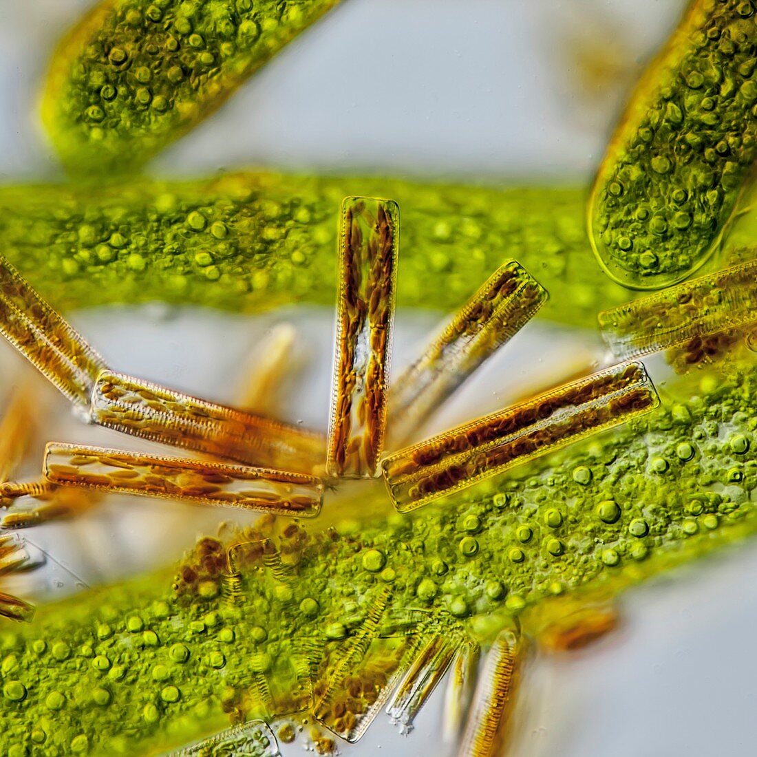
| Light micrograph of Cladophora sp. (Gr. klados,branch & Gr. phoras,bearing) filaments with attached Diatoma sp. freshwater diatoms. The cells of Diatoma are attached together by means of mucilage pads. Diatoma cells are characterized by the brownish chloroplasts in contrast to the green chloroplast of Cladophora. Microscopic contrast method : Differential interference contrast. Magnification 380x when printed at 10 centimetres wide | |
| Licence : | Droits gérés |
| Crédit: | Science Photo Library / Guenther, Gerd |
| Taille de l’image : | 4180 px × 4180 px |
| Model Release : | Non requis |
| Property Release : | Non requis |
| Restrictions : | - |
Prix pour cette image À partir de 45 €
Produit vendu
(Calendrier, Carte postale, Carte de vœux, Impression sur textile, Packaging etc)
À partir de 45 €
Usage commercial
(Affichage, Annonce presse, Annonce TV, Carte, Digital - hors rés. sociaux, Digital - rés. sociaux etc)
À partir de 45 €
Éditorial
(Digital, Journal, Livre, Livre pratique, Magazine, Télévision etc)
À partir de 60 €
Usage non-commercial
(Digital - hors rés. sociaux, Digital - rés. sociaux etc)
À partir de 120 €
Mots clés
- alga,
- algae,
- algologie,
- algue,
- algue verte,
- algues,
- aquatique,
- aucun,
- biologie,
- biologique,
- botanique,
- CLADOPHORA,
- contraste d'interférence différentielle,
- contraste interférentiel différentiel,
- contratse différentiel d'interférence,
- diatomée,
- eau douce,
- flore,
- micro-organisme,
- microscope optique,
- microscopie optique,
- monocellulaire,
- nature,
- personne,
- phycologie,
- phycologique,
- unicellulaire,
- vie d'un étang,
- vie primitive