Fetal testis,light micrograph
Numéro d’image : 11723327
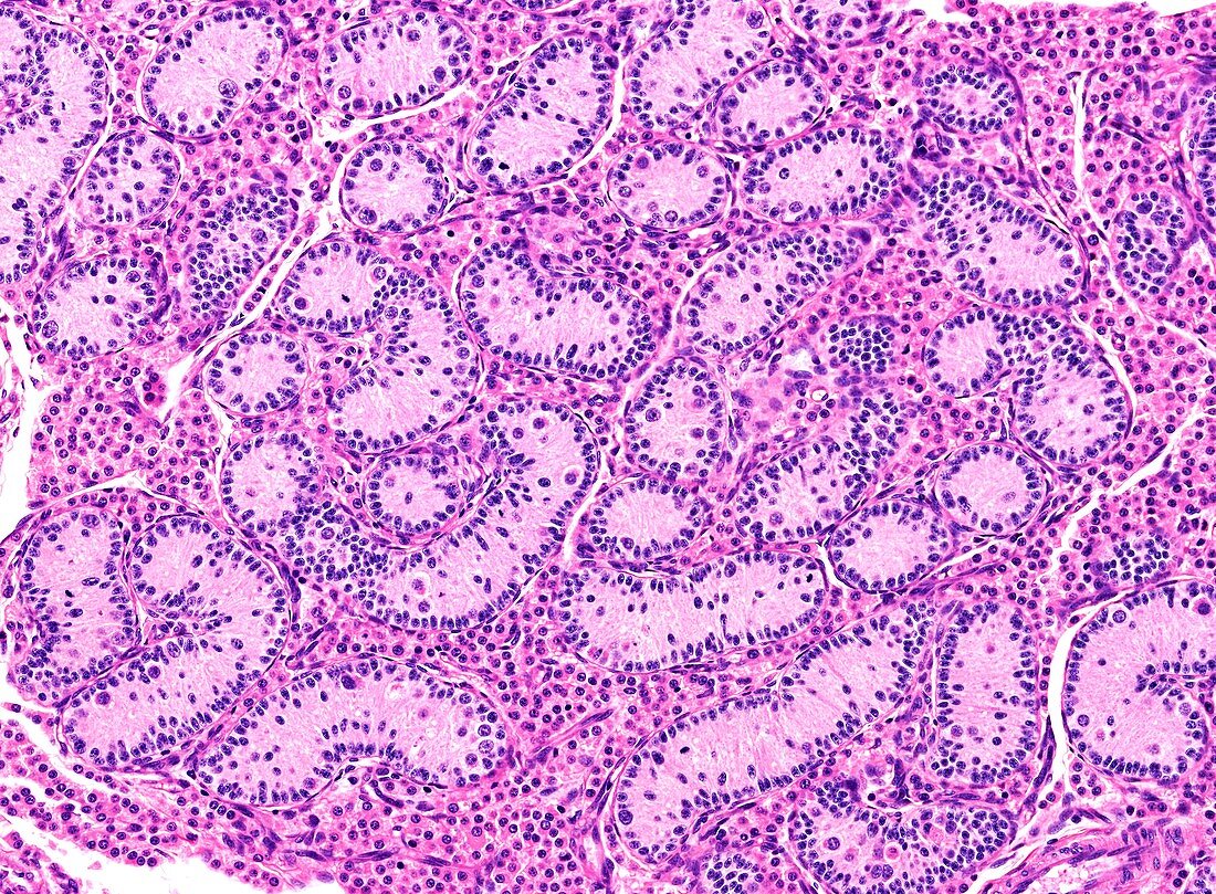
| Light microscopy of a fetal testis. The characteristic features are i) the solid seminiferous cords that in postnatal life will become seminiferous tubules,and ii) the relatively high proportion of Leydig cells between the cords which will diminish in quantity in postnatal life. The cords contain mostly Sertoli cells and fewer spermatogonia (round cells). The spermatogonia will become the stem cells for subsequent sperm development during spermatogenesis. Magnification x200 when printed at 10 cm height | |
| Licence : | Droits gérés |
| Crédit: | Science Photo Library / Microscape |
| Taille de l’image : | 4875 px × 3585 px |
| Model Release : | Non requis |
| Property Release : | Non requis |
| Restrictions : | - |
Prix pour cette image À partir de 45 €
Produit vendu
(Calendrier, Carte postale, Carte de vœux, Impression sur textile, Packaging etc)
À partir de 45 €
Usage commercial
(Affichage, Annonce presse, Annonce TV, Carte, Digital - hors rés. sociaux, Digital - rés. sociaux etc)
À partir de 45 €
Éditorial
(Digital, Journal, Livre, Livre pratique, Magazine, Télévision etc)
À partir de 60 €
Usage non-commercial
(Digital - hors rés. sociaux, Digital - rés. sociaux etc)
À partir de 120 €
Mots clés
- biologie,
- biologique,
- catégorie,
- cellule de germe,
- cellule de Leydig,
- cellule de Sertoli,
- cellule germinale,
- coupe,
- épithélium séminifère,
- gonade,
- histologie,
- histologique,
- microscope optique,
- microscope photonique,
- microscopie optique,
- microscopie photonique,
- partie,
- reproduction,
- reproduction masculine,
- section,
- spermatogenèse,
- spermatozoïde,
- système reproducteur masculin,
- système reproductif,
- testicules,
- testis,
- tube séminifère